
Description
Overview
The FireGene Mouse Brain Dissociation Kit is engineered to produce high-quality single-cell suspensions from mouse brain tissue using an optimized enzymatic process. This kit is ideal for neuroscience research, enabling precise gene expression profiling and functional studies at the single-cell level.
Background Information
-
Driven by Clinical and Scientific Research Needs:
-
Increased demand for high-resolution studies of cellular heterogeneity in the brain.
- Traditional methods fail to preserve delicate brain cell populations and often yield low viability.
-
Single-cell sequencing of mouse brain tissue enables:
-
Detailed gene expression analysis.
-
Functional characterization of neurons, glia, and other CNS cell types.
-
Discovery of new biomarkers and therapeutic targets for brain disorders.
-
-
Critical for exploring neurodevelopment, neurodegeneration, and brain repair mechanisms.
-
-
Background of Technological Development:
-
Overcomes limitations of mechanical and chemical dissociation methods.
-
Employs advanced enzymatic protocols that:
-
Use optimized enzyme concentrations and incubation times.
-
Minimize damage to fragile neural cells.
-
Preserve cell viability and diversity.
-
-
Designed to ensure consistent, reproducible dissociation outcomes.
-
Detection Principle
-
Utilizes a synergistic enzymatic digestion method optimized for brain tissue.
-
Process highlights:
-
Mouse brain tissue is cut into small pieces for uniform exposure.
-
Sequential addition of enzyme mixtures facilitates gentle tissue breakdown.
-
Incubation is performed under controlled temperature and timing conditions.
-
Enzymes selectively degrade the extracellular matrix and cell junctions.
-
-
Final output:
-
A clean, high-viability single-cell suspension ready for scRNA-seq, flow cytometry, and downstream analysis.
-
Specifications
| Applications | Single-cell sequencing, cell culture or other cell-related detections |
| Compatible Sample Types | Mouse brain tissue |
| Supported Instruments | Water bath, horizontal centrifuge, cell counter |
| Storage | -20 °C / 4 °C |
| Shelf-life | 24 months at -20 °C 12 months at 4 °C |
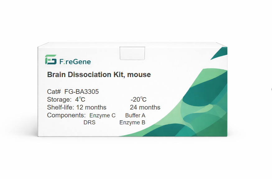
Kit Components
10 reactions
| Component | 10 Tests/Kit | Storage | Shelf-life |
|---|---|---|---|
| Buffer A | 1*21.6 mL | -20 °C | 24 months |
| Enzyme B | 1*8 mL | -20 °C | 24 months |
| Enzyme C | 1*100 μL | 4 °C | 12 months |
| DRS(Mouse Brain Cell Debris Removal Buffer) | 1*10 mL | 4 °C | 12 months |
50 reactions
| Component | 10 Tests/Kit | Storage | Shelf-life |
|---|---|---|---|
| Buffer A | 5*21.6 mL | -20 °C | 24 months |
| Enzyme B | 5*8 mL | -20 °C | 24 months |
| Enzyme C | 5*100 μL | 4 °C | 12 months |
| DRS(Mouse Brain Cell Debris Removal Buffer) | 5*10 mL | 4 °C | 12 months |
Product Q&A
1. Q: Is this kit only suitable for mouse brain tissue? Is it applicable to brain tissue of other animals such as rats and rabbits? Can it be used for non-brain tissues of mice (e.g., liver, kidney)?
A: This kit is specially designed for mouse brain tissue and is only suitable for the dissociation of mouse brain tissue. It is not recommended for use with brain tissue of other animals like rats and rabbits for the time being. The cell density and extracellular matrix composition of brain tissue vary among different animals, and the enzyme ratio and buffer components in the kit are optimized for mouse brain tissue. Using it on other animals may result in low dissociation efficiency or poor cell viability. Meanwhile, it cannot be used for non-brain tissues of mice. Non-brain tissues (such as liver and kidney) have significantly different structures from brain tissue, so a dedicated dissociation kit for the corresponding tissue (e.g., FG-BA3323 Liver Tissue Dissociation Kit) should be used.
2. Q: The components of the kit have different storage conditions. What should be noted before mixing and using them? If Enzyme C is accidentally stored frozen at -20°C, can it still be used?
A: Two points should be noted before mixing and using: First, confirm that all components are within their validity period and stored in compliance with the required conditions (Buffer A and Enzyme B at -20°C; Enzyme C and DRS at 4°C in the dark). Second, before use, take Buffer A and Enzyme B out of the -20°C freezer, thaw them at room temperature, and mix thoroughly. Enzyme C and DRS can be directly taken out of the 4°C refrigerator and mixed well, with repeated freezing and thawing avoided. If Enzyme C is accidentally stored frozen at -20°C, it cannot be used anymore. Enzyme C is an enzyme preparation, and low-temperature freezing will destroy its spatial structure, leading to complete loss of enzyme activity. Using it will fail to dissociate the tissue effectively, and you need to contact the manufacturer to purchase a replacement.
3. Q: Step 1 requires cutting about 200mg of fresh tissue. What impact will insufficient tissue quantity (e.g., only 50mg) or excessive tissue quantity (e.g., 300mg) have on the dissociation effect? How to adjust the operation?
A: Insufficient tissue quantity (50mg) will lead to a relative excess of enzyme reagents, which may cause over-digestion of cells and reduce cell viability. Excessive tissue quantity (300mg) will result in insufficient enzyme reagents, which cannot fully break down the extracellular matrix, leading to incomplete tissue dissociation, a large number of tissue clumps, and low single-cell yield. Adjustment methods: When the tissue quantity is insufficient, the dosage of each reagent can be reduced proportionally (e.g., Buffer A reduced to 540μL, Enzyme B to 200μL, Enzyme C to 2.5μL) to ensure the ratio of enzyme to tissue is appropriate. When the tissue quantity is excessive, it should be divided into two portions for processing, and each portion should be operated with the reagent dosage corresponding to 200mg of tissue to avoid insufficient reagents affecting dissociation.
4. Q: In Step 3, the digestion time is 20-30 minutes, and quality inspection is required every 3-5 minutes. What is the specific operation of quality inspection? How to judge whether digestion needs to be stopped?
A: Specific operation of quality inspection: Take 10μL of cell suspension, add 10μL of AOPI or trypan blue staining solution (self-prepared), mix well, drop it onto a cell counting plate, and observe under a cell counter or microscope. There are two criteria for judging whether to stop digestion: First, cell viability. If the viability is low, digestion must be stopped even if the tissue is not fully dissociated to avoid over-digestion. Second, cell dispersion. When the proportion of tissue clumps in the field of view is ≤5% and the proportion of single cells is ≥90%, digestion can be stopped. If there are still many tissue clumps after 30 minutes but the cell viability is ≥70%, digestion can be extended for 5 minutes, but quality inspection must be conducted again to avoid exceeding 35 minutes.
5. Q: Both Step 5 and Step 6 require filtration with a 70μm cell sieve. Why does Step 6 require rinsing the original centrifuge tube and filtering again? What consequences will occur if the rinsing and filtering in Step 6 are omitted?
A: The purpose of rinsing and filtering in Step 6 is to recover the cells remaining on the wall of the original centrifuge tube. Digested cells may adhere to the inner wall of the centrifuge tube, and filtering only in Step 5 will cause the loss of these cells, reducing the single-cell yield. If this step is omitted, the cell yield may decrease by 15%-20%. Especially for samples with a small initial tissue quantity, it will seriously affect subsequent experiments (e.g., single-cell sequencing requires a sufficient number of cells). Therefore, the tube wall must be rinsed with 3mL of RPMI 1640 medium or PBS containing 5% FBS, and the rinsing solution must be filtered through the same 70μm cell sieve to ensure full cell recovery.
6. Q: After adding DRS in Step 11, it is necessary to gently pipette 10 times with a 5mL pipette, and "vortex oscillation is not allowed". What problems will vortex oscillation cause? What impacts will insufficient or excessive pipetting times have?
A: Vortex oscillation will generate strong mechanical force, which damages the cell membrane of mouse brain cells, leading to cell rupture. At the same time, it will cause uneven mixing of DRS and cell suspension, affecting the subsequent density gradient stratification effect. Insufficient pipetting times (less than 10 times) will result in insufficient mixing of DRS and cell suspension, making it impossible to form stable debris layers and cell layers after centrifugation, leading to incomplete debris removal. Excessive pipetting times (more than 15 times) will damage cells due to mechanical friction, increase the proportion of dead cells, and may disrupt the density system of DRS, also affecting stratification. It is necessary to strictly control the pipetting to 10 times, with gentle movements to avoid generating air bubbles.
7. Q: Step 12 requires slowly adding 3mL of PBS along the tube wall to "gently cover the top layer, and never mix". If mixing occurs accidentally during addition, how to handle it? Is it necessary to re-operate?
A: If mixing occurs accidentally when adding PBS, it will destroy the density gradient foundation formed by DRS and cell suspension. After centrifugation, it will be impossible to clearly separate the debris layer, supernatant, and cell pellet, resulting in the failure of debris removal. Therefore, re-operation is mandatory. For re-operation, centrifuge the mixed solution at 300×g for 5 minutes at 4°C, discard the supernatant, resuspend the pellet with 2mL of PBS containing 5% FBS, then add 1mL of DRS and gently pipette 10 times according to Step 11. After that, slowly add 3mL of PBS along the tube wall again, ensuring no mixing to avoid re-occurrence of errors.
8. Q: Steps 13 and 14 emphasize "a horizontal centrifuge must be used" and "the centrifuge tube must be handled gently". What impact will using a vertical centrifuge or handling the tube roughly after centrifugation have on stratification?
A: When a vertical centrifuge is used, the direction of centrifugal force is perpendicular to the centrifuge tube, making it impossible for the solution to be evenly stratified according to the density gradient. This will cause debris, cells, and PBS to mix, and no clear three-layer structure can be separated. Rough handling of the centrifuge tube after centrifugation will cause violent shaking of the solution inside the tube, and the formed three-layer structure (supernatant, debris layer, cell pellet) will mix with each other. Debris will re-mix into the cell pellet, resulting in the loss of debris removal effect. Therefore, a horizontal centrifuge must be used, and after centrifugation, the centrifuge tube should be slowly taken out with both hands supporting the bottom to avoid any violent shaking.
9. Q: When adding red blood cell lysis buffer (FG-BA3311) in Step 15, what volume is the "three times the volume" calculated based on? Can the red blood cell lysis time on ice (5 minutes) be shortened or extended?
A: "Three times the volume" is calculated based on the "appropriate volume of PBS containing 5% FBS" in Step 15. For example, if 100μL of PBS containing 5% FBS is used to resuspend the pellet, 300μL of red blood cell lysis buffer should be added. The red blood cell lysis time on ice cannot be shortened or extended: Shortening it to less than 5 minutes will result in incomplete red blood cell lysis, and residual red blood cells will mix into the brain cell suspension, interfering with subsequent cell counting and experiments (e.g., non-specific signals of red blood cells in flow cytometry analysis). Extending it to more than 5 minutes will make the lysis buffer toxic to brain cells, leading to decreased brain cell viability, especially for sensitive neuron cells. Thus, the time must be strictly controlled at 5 minutes.
10. Q: After quality control in Step 21, it is required to "carry out subsequent experiments immediately". If subsequent experiments cannot be conducted immediately, can the prepared brain cell suspension be stored for a short period? What are the restrictions on storage conditions and time?
A: Short-term storage is possible, but there are strict restrictions on storage conditions and time: The cell suspension needs to be adjusted to a concentration of 1×10⁶-1×10⁷ cells/mL (using PBS containing 5% FBS), placed in a sterile low-adhesion centrifuge tube, sealed, and stored in a 4°C refrigerator. The storage time should not exceed 1 hour, and repeated shaking should be avoided during storage. If stored for more than 1 hour, the viability of brain cells will decrease significantly (may be lower than 60%), and a small amount of new cell debris will appear. If stored for more than 2 hours, the cells will basically lose their viability and cannot be used for subsequent experiments (e.g., cell culture, single-cell sequencing). Before use, trypan blue staining must be re-conducted for quality inspection, and only cells with viability ≥70% can be used.
Files Download Links