
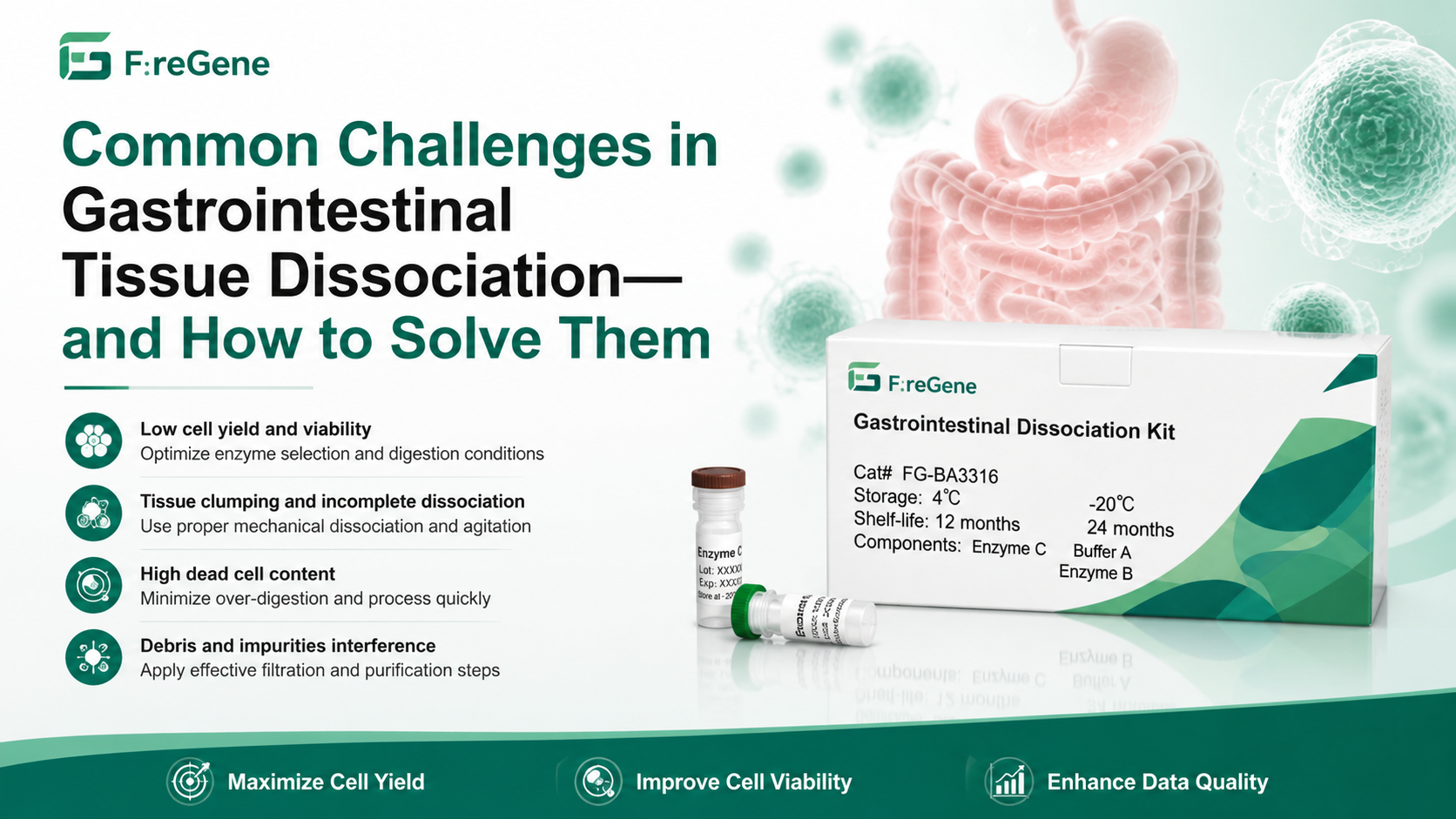
Common Challenges in Gastrointestinal Tissue Dissociation—and How to Solve Them
Introduction
Over the past decade, gastrointestinal research has undergone a remarkable transformation. Instead of analyzing tissues as homogeneous populations, researchers can now investigate the molecular characteristics of individual cells using technologies such as single-cell RNA sequencing (scRNA-seq), single-cell ATAC-seq, spatial transcriptomics, CyTOF, high-dimensional flow cytometry, multiplex imaging, and organoid culture.
These technologies have fundamentally changed our understanding of gastrointestinal diseases.
Scientists are now uncovering previously unknown epithelial stem cell populations, identifying rare immune subsets associated with inflammatory bowel disease (IBD), mapping the tumor microenvironment of colorectal cancer, and revealing complex interactions between intestinal epithelial cells, stromal cells, immune cells, and the gut microbiome.
Large-scale international projects—including the Human Cell Atlas and multiple gastrointestinal cell atlas initiatives—have generated millions of single-cell datasets that continue to reshape digestive disease research.
However, regardless of how advanced the downstream technology becomes, every successful experiment depends on one critical upstream step:
Obtaining a high-quality single-cell suspension.
Unfortunately, this is often easier said than done.
Unlike tissues such as spleen or peripheral blood, gastrointestinal tissue presents exceptional technical challenges during dissociation. Researchers frequently encounter problems including:
- Low cell viability
- Excessive cell death
- Cell clumping
- Poor epithelial recovery
- RNA degradation
- Over-digestion
- Batch-to-batch variability
- Stress-induced transcriptional artifacts
Even small differences during tissue digestion can significantly alter downstream sequencing results. Poor-quality cell suspensions often lead to lower sequencing depth, increased ambient RNA contamination, reduced cell recovery, inaccurate clustering, and misleading biological conclusions.
In many cases, researchers mistakenly attribute poor sequencing quality to library preparation or sequencing platforms when the true source of the problem originates much earlier—during tissue dissociation.
As gastrointestinal research increasingly moves toward multiomics, spatial biology, and AI-assisted data analysis, the importance of standardized tissue preparation continues to grow.
This article reviews the most common challenges encountered during gastrointestinal tissue dissociation, explains why these problems occur, and provides practical solutions that can help researchers generate highly viable, reproducible single-cell suspensions suitable for modern downstream applications.
Why Gastrointestinal Tissue Is One of the Most Difficult Tissues to Dissociate
Many researchers assume that tissue dissociation simply involves incubating tissue fragments with collagenase until cells are released.
In reality, gastrointestinal tissue is one of the most structurally complex organs in the human body.
Its unique architecture has evolved to perform digestion, nutrient absorption, immune surveillance, microbial regulation, and continuous epithelial renewal simultaneously. While these biological functions are essential in vivo, they create numerous obstacles during laboratory tissue processing.
Understanding these structural challenges is the first step toward selecting an appropriate dissociation strategy.
1. A Dense and Complex Extracellular Matrix
The gastrointestinal extracellular matrix (ECM) differs substantially from many other tissues.
Instead of containing only loose connective tissue, the intestinal wall includes a highly organized network of:
- Collagen I
- Collagen III
- Collagen IV
- Fibronectin
- Laminin
- Elastin
- Proteoglycans
- Hyaluronic acid
These ECM components form a robust scaffold that tightly anchors epithelial cells, fibroblasts, endothelial cells, smooth muscle cells, and immune populations.
While this architecture maintains tissue integrity in vivo, it also makes enzymatic digestion considerably more difficult.
Insufficient digestion leaves large tissue fragments intact, whereas excessive digestion damages delicate epithelial populations and alters gene expression.
Finding the correct balance between digestion efficiency and cell preservation is therefore one of the greatest challenges in gastrointestinal tissue processing.
2. The Protective Mucus Barrier
Unlike most solid organs, the gastrointestinal tract is covered by a thick mucus layer composed primarily of mucins secreted by goblet cells.
This mucus performs several essential biological functions:
- Protects epithelial cells from mechanical damage
- Prevents bacterial invasion
- Maintains microbiome homeostasis
- Traps digestive enzymes
- Lubricates intestinal contents
From a tissue dissociation perspective, however, mucus creates significant technical complications.
The highly viscous mucus can:
- Trap liberated cells before they enter suspension
- Reduce enzyme penetration into tissue fragments
- Increase cell aggregation
- Interfere with filtration
- Promote clogging during flow cytometry or single-cell library preparation
Residual mucus is one of the most common yet frequently overlooked causes of cell clumping and poor single-cell recovery.
3. Multiple Tissue Layers Require Different Digestion Conditions
The gastrointestinal tract is not a single tissue but rather a highly organized multilayered organ composed of distinct anatomical regions.
Each layer has unique structural properties and enzyme sensitivities:
- Mucosa
- Lamina propria
- Muscularis mucosae
- Submucosa
- Circular muscle
- Longitudinal muscle
- Serosa
Furthermore, the stomach, small intestine, colon, and rectum each contain different extracellular matrix compositions, cellular architectures, and connective tissue densities.
A digestion protocol that performs well for mouse small intestine may produce poor results when applied to human colon samples.
Similarly, inflammatory tissues often require different digestion strategies than healthy tissues due to fibrosis, immune infiltration, and extracellular matrix remodeling.
This variability explains why universal digestion protocols rarely produce optimal results across all gastrointestinal specimens.
4. Rapid Epithelial Cell Turnover Creates Fragile Cell Populations
The intestinal epithelium is among the fastest-renewing tissues in the human body.
Most epithelial cells are replaced every three to five days.
Continuous regeneration depends on intestinal stem cells located at the base of the crypts, which differentiate into specialized cell types including:
- Enterocytes
- Goblet cells
- Paneth cells
- Enteroendocrine cells
- Tuft cells
- M cells
Although biologically fascinating, these rapidly proliferating cells are extremely sensitive to mechanical stress and prolonged enzymatic digestion.
Excessive pipetting, overly concentrated enzyme mixtures, or extended incubation times can selectively damage epithelial populations while leaving more resilient stromal or immune cells relatively unaffected.
As a result, researchers may unknowingly generate datasets that underrepresent critical epithelial cell populations, introducing substantial bias into downstream analyses.
5. High Cellular Diversity Increases Dissociation Complexity
Few organs contain the extraordinary cellular diversity found within gastrointestinal tissue.
A typical intestinal biopsy may include:
- Epithelial cells
- Fibroblasts
- Endothelial cells
- Smooth muscle cells
- Macrophages
- Monocytes
- Dendritic cells
- B cells
- Plasma cells
- CD4⁺ T cells
- CD8⁺ T cells
- Regulatory T cells
- Innate lymphoid cells
- Neurons
- Glial cells
Each cell type differs in size, membrane composition, mechanical resistance, enzyme sensitivity, and susceptibility to apoptosis.
Consequently, no single digestion condition is equally suitable for every population.
Protocols optimized for immune cell recovery may compromise epithelial viability, while methods designed to maximize epithelial yield may underrepresent stromal or immune compartments.
The challenge is therefore not simply releasing the largest number of cells—it is preserving the original cellular composition of the tissue as faithfully as possible.
Why Tissue Dissociation Matters More Than Ever
The quality of tissue dissociation influences virtually every downstream analytical platform.
For example:
- In single-cell RNA sequencing, poor dissociation reduces viable cell recovery, increases ambient RNA contamination, and introduces stress-response gene signatures.
- In flow cytometry, cell aggregates and debris can clog instruments and reduce sorting efficiency.
- In organoid culture, damaged stem cells fail to establish long-term cultures.
- In spatial transcriptomics, dissociation optimization often informs parallel workflows used to validate spatially identified cell populations.
As researchers increasingly integrate multiomics, spatial biology, and AI-assisted computational analysis, the demand for standardized, reproducible tissue preparation workflows has never been greater.
This is one reason why many laboratories are transitioning away from generic enzyme cocktails toward tissue-specific solutions such as the FireGene Gastrointestinal Dissociation Kit, which is specifically optimized for efficient digestion of gastrointestinal tissues while preserving cell viability and broad cellular representation.
Common Challenges in Gastrointestinal Tissue Dissociation—and How to Solve Them
Challenge 1: Low Cell Viability—The Most Common Cause of Failed Single-Cell Experiments
Among all tissue dissociation problems, low cell viability is undoubtedly the most frequently reported issue in gastrointestinal research. Whether the downstream application is single-cell RNA sequencing, flow cytometry, organoid establishment, or primary cell culture, excessive cell death at the dissociation stage can compromise the entire experiment.
In many laboratories, researchers focus primarily on maximizing cell yield. While recovering a large number of cells may appear desirable, high yield alone does not guarantee high-quality data. A suspension containing millions of damaged or dying cells is often less valuable than a smaller population of highly viable cells that accurately represents the original tissue.
For most single-cell sequencing platforms, cell viability above 85% is generally recommended before library preparation, while many researchers aim for 90–95% viability whenever possible. Once viability drops below acceptable levels, the consequences extend far beyond simple cell loss.
What Happens When Cell Viability Is Poor?
Low viability affects nearly every stage of downstream analysis.
Common consequences include:
- Increased ambient RNA contamination
- Reduced unique molecular identifier (UMI) counts
- Lower gene detection per cell
- Higher mitochondrial RNA percentages
- Greater proportions of apoptotic cells
- Reduced recovery of fragile epithelial populations
- Increased sequencing costs due to unusable libraries
Dead cells release intracellular RNA into the surrounding solution. During droplet encapsulation, this extracellular RNA may be captured together with intact cells, producing misleading transcriptional profiles that do not accurately reflect biological reality.
This phenomenon, commonly referred to as ambient RNA contamination, can create artificial gene expression signatures that complicate downstream clustering and cell-type annotation.
Why Does Viability Drop During Gastrointestinal Tissue Dissociation?
Several factors often contribute simultaneously.
1. Over-Digestion
The most common mistake is assuming that longer digestion always produces better cell recovery.
In reality, prolonged enzyme exposure gradually damages cell membranes, disrupts cell-cell junctions, and activates apoptotic pathways.
Fragile epithelial stem cells are usually affected first, followed by differentiated epithelial populations.
Ironically, extending digestion beyond the optimal endpoint often decreases the total number of usable cells.
2. Excessive Mechanical Force
Mechanical disruption is necessary to separate partially digested tissue.
However, aggressive pipetting, vigorous vortexing, or repeated aspiration through narrow pipette tips generates substantial shear stress.
These forces can rupture plasma membranes and selectively destroy larger epithelial cells while leaving smaller immune cells relatively intact.
The resulting suspension may appear highly cellular, but its composition no longer reflects the original tissue.
3. Delayed Tissue Processing
Time is one of the most overlooked variables.
Immediately after surgical removal or biopsy collection, tissues experience ischemia.
Without oxygen and nutrient supply, cells begin activating stress-response pathways.
Delays of even one to two hours may significantly reduce epithelial viability.
Whenever possible, tissue should be processed immediately after collection.
4. Temperature Fluctuations
Temperature control is equally important.
Digestive enzymes require physiological temperatures for efficient activity.
However, cells should remain on ice before digestion and immediately after enzyme neutralization.
Repeated warming and cooling cycles increase cellular stress and accelerate RNA degradation.
5. Incorrect Enzyme Selection
Many laboratories still rely on generic collagenase formulations originally developed for other tissues.
Unfortunately, gastrointestinal tissue contains unique extracellular matrix components and mucus barriers that require carefully balanced enzyme combinations.
Using inappropriate enzymes often forces researchers to compensate by increasing incubation time, which ultimately reduces viability.
Practical Strategies to Improve Cell Viability
Improving viability rarely depends on a single modification.
Instead, success comes from optimizing the entire workflow.
Standardize Tissue Size Before Digestion
Large tissue fragments digest unevenly.
The outer surface becomes over-digested while the interior remains largely intact.
Cutting tissue into uniformly sized pieces dramatically improves enzyme penetration and shortens digestion time.
Optimize Digestion Rather Than Maximizing Digestion
A useful principle is:
Stop digestion when cells are being released efficiently—not when every tissue fragment has disappeared.
Leaving a few small tissue fragments is often preferable to exposing liberated cells to unnecessary enzyme activity.
Minimize Mechanical Stress
Instead of vigorous pipetting:
- Use wide-bore pipette tips
- Mix gently every few minutes
- Avoid vortexing whenever possible
Mechanical disruption should assist enzymatic digestion rather than replace it.
Maintain Physiological Conditions
Successful protocols typically include:
- Immediate tissue transport in cold preservation buffer
- Rapid transfer to digestion solution
- Controlled incubation at recommended temperatures
- Immediate enzyme neutralization
- Cold handling during all post-digestion steps
Use Gastrointestinal-Specific Dissociation Reagents
Perhaps the simplest way to improve viability is selecting reagents specifically optimized for gastrointestinal tissue.
Unlike generic collagenase mixtures, tissue-specific enzyme formulations are designed to efficiently digest intestinal extracellular matrix while minimizing unnecessary cellular damage.
For researchers routinely performing intestinal single-cell studies, using an optimized workflow such as the FireGene Gastrointestinal Dissociation Kit can significantly simplify protocol optimization while improving cell viability across epithelial, stromal, and immune populations.
Challenge 2: Cell Clumping—A Hidden Obstacle That Reduces Data Quality
Few problems are as frustrating as preparing what appears to be an excellent single-cell suspension, only to discover large cell aggregates during counting or instrument loading.
Cell clumping is particularly common in gastrointestinal tissues because of their abundant extracellular matrix, high mucus content, and large numbers of dying cells released during digestion.
Although aggregates may seem like a minor inconvenience, they can dramatically affect downstream applications.
For droplet-based single-cell sequencing platforms such as 10x Genomics Chromium, cell aggregates increase the probability of capturing multiple cells within a single droplet. These doublets or multiplets can generate hybrid transcriptomes that are difficult to identify computationally and may lead to false cell populations during clustering.
For flow cytometry and fluorescence-activated cell sorting (FACS), aggregates can clog fluidics systems, reduce acquisition speed, increase abort rates, and compromise sorting purity.
Why Do Cell Clumps Form?
Cell aggregation is rarely caused by a single factor. Instead, several biological and technical mechanisms often work together.
Extracellular DNA Released from Dead Cells
When cells rupture during digestion, fragmented genomic DNA is released into the suspension.
DNA is highly viscous and acts like a biological adhesive, causing neighboring cells to stick together.
Even relatively small numbers of dead cells can generate surprisingly large aggregates.
This is why DNase is included in many optimized tissue dissociation protocols.
Residual Mucus
The gastrointestinal tract naturally produces large quantities of mucins.
Incomplete removal of mucus increases suspension viscosity and traps individual cells within sticky mucus networks.
Researchers often mistake these mucus-associated aggregates for incomplete digestion when the actual problem is inadequate mucus disruption.
Incomplete Extracellular Matrix Digestion
If collagen fibers remain only partially digested, liberated cells remain physically connected through extracellular matrix remnants.
Microscopic examination typically reveals irregular tissue fragments rather than free-floating individual cells.
Increasing mechanical force is rarely the correct solution.
Instead, enzyme optimization generally produces better results.
Excessive Centrifugation
High centrifugation speeds compress cells into dense pellets.
Repeated resuspension may not fully separate tightly packed aggregates.
Using moderate centrifugal forces helps preserve single-cell suspensions.
How to Recognize Cell Clumping Early
Rather than waiting until sequencing fails, researchers should evaluate suspension quality immediately after dissociation.
Typical warning signs include:
- Visible strands during pipetting
- Uneven cell distribution
- Rapid sedimentation
- Large aggregates under the microscope
- Inconsistent automated cell counter results
- Frequent instrument clogging
Routine microscopic examination remains one of the simplest and most valuable quality-control steps.
Strategies to Reduce Cell Aggregation
Several practical approaches can substantially improve suspension quality.
Include DNase During Digestion
DNase breaks down extracellular DNA released by damaged cells, reducing suspension viscosity and preventing DNA-mediated aggregation.
Perform Sequential Filtration
Filtering through progressively smaller strainers—such as 100 μm, followed by 70 μm and finally 40 μm—removes residual tissue fragments while minimizing mechanical stress.
Sequential filtration is generally more effective than forcing samples through a single fine mesh.
Avoid Excessive Pipetting
Ironically, aggressive attempts to disperse aggregates often create additional dead cells, releasing even more extracellular DNA.
Gentle handling usually produces better long-term results.
Remove Dead Cells Before Library Preparation
If significant debris remains after digestion, incorporating a dead-cell removal step can substantially improve downstream sequencing performance.
Removing apoptotic cells not only reduces aggregation but also decreases ambient RNA contamination and improves overall library quality.
Challenge 3: Poor Recovery of Fragile Epithelial Cells—Why Your Most Important Cells May Be Missing
For many gastrointestinal studies, epithelial cells are the primary biological focus. Whether investigating intestinal stem cell biology, inflammatory bowel disease (IBD), colorectal cancer, host-microbiome interactions, or organoid development, researchers rely on accurate representation of epithelial populations to generate meaningful biological insights.
Unfortunately, epithelial cells are also among the most vulnerable cell types during tissue dissociation.
Unlike immune cells, which often exist as relatively independent cells within the lamina propria, epithelial cells are tightly organized into a continuous barrier through adherens junctions, tight junctions, and desmosomes. They are firmly anchored to the basement membrane and surrounded by a highly specialized extracellular matrix. Releasing these cells without damaging them requires careful optimization of both enzymatic and mechanical dissociation.
As a result, many researchers unknowingly generate datasets that contain abundant immune and stromal cells but significantly underrepresent epithelial populations. This imbalance may not be obvious during cell counting, yet it can profoundly influence downstream biological interpretation.
Why Are Epithelial Cells So Easily Lost?
Several biological characteristics make epithelial cells particularly susceptible to dissociation-induced damage.
Tight Cell–Cell Junctions
The intestinal epithelium functions as a selective barrier that regulates nutrient absorption while preventing microbial invasion. To maintain this barrier, neighboring epithelial cells are connected by an extensive network of tight junction proteins, adherens junctions, and desmosomes.
These structures are mechanically robust in vivo but present a major challenge during tissue dissociation. Breaking these junctions often requires enzymatic digestion combined with gentle mechanical disruption. Excessive force, however, may tear cells apart rather than separating them cleanly.
Strong Attachment to the Basement Membrane
Unlike immune cells that migrate freely within connective tissue, epithelial cells remain firmly attached to the basement membrane through integrins and other adhesion molecules.
Incomplete digestion often leaves epithelial sheets attached to tissue fragments, causing researchers to discard valuable cell populations during filtration.
High Metabolic Activity
Intestinal epithelial cells are metabolically active because they continuously absorb nutrients and undergo rapid turnover.
This high metabolic demand also makes them particularly sensitive to:
- Oxygen deprivation
- Temperature fluctuations
- Oxidative stress
- Mechanical injury
- Prolonged enzyme exposure
Even relatively short processing delays can trigger apoptosis in these cells.
Certain Epithelial Cell Types Are Especially Vulnerable
Not all epithelial populations respond equally during tissue dissociation.
Intestinal Stem Cells
Located at the base of intestinal crypts, stem cells drive continuous epithelial renewal.
Because they are embedded deep within crypt structures and surrounded by supporting niche cells, they require efficient—but gentle—matrix digestion for successful recovery.
Harsh digestion protocols frequently reduce stem cell recovery, limiting downstream organoid formation and stem-cell transcriptomic analyses.
Goblet Cells
Goblet cells synthesize and secrete mucins that form the protective mucus barrier.
Ironically, the same mucus they produce also complicates tissue dissociation.
Goblet cells often rupture during excessive mechanical mixing, releasing additional mucins that further increase suspension viscosity and promote aggregation.
Paneth Cells
Paneth cells reside adjacent to stem cells within intestinal crypts.
They contain abundant antimicrobial granules that are highly sensitive to mechanical damage.
Suboptimal digestion frequently leads to poor Paneth cell representation in single-cell datasets.
Enteroendocrine Cells
Although relatively rare, enteroendocrine cells regulate numerous gastrointestinal hormones.
Because these cells represent only a small fraction of total epithelial populations, even modest cell loss may eliminate them entirely from sequencing datasets.
Tuft Cells
Tuft cells have become increasingly important in studies of immunity and host-parasite interactions.
However, their rarity makes them particularly vulnerable to dissociation bias.
Failure to recover tuft cells may lead researchers to incorrectly conclude that these populations are absent.
Signs That Epithelial Recovery Is Poor
Researchers should monitor several indicators during quality control.
Warning signs include:
- Unexpectedly high proportions of immune cells
- Very low EPCAM-positive populations
- Reduced expression of epithelial marker genes
- Missing crypt-associated cell clusters
- Poor organoid establishment
- Low epithelial diversity in scRNA-seq datasets
Importantly, these issues may not reflect biological differences between samples—they often result from technical variation introduced during dissociation.
How to Improve Epithelial Cell Recovery
Successful epithelial isolation depends on balancing efficient tissue digestion with gentle cell handling.
Recommended practices include:
- Standardizing tissue fragment size before digestion
- Avoiding prolonged incubation
- Using gentle pipetting techniques
- Maintaining appropriate enzyme concentrations
- Neutralizing enzymes promptly
- Keeping samples cold during post-digestion processing
Equally important is the use of gastrointestinal-specific dissociation reagents that have been optimized to preserve delicate epithelial populations while efficiently releasing stromal and immune cells.
Researchers working with intestinal organoids, epithelial regeneration, colorectal cancer, or inflammatory bowel disease often benefit from using optimized workflows such as the FireGene Gastrointestinal Dissociation Kit, which is designed to maximize epithelial recovery while maintaining high overall cell viability.
Challenge 4: Immune Cell Bias—When Your Dataset No Longer Represents the Original Tissue
One of the least discussed—but perhaps most important—problems in gastrointestinal tissue dissociation is cell-type bias.
Researchers often assume that every recovered cell accurately reflects the original tissue composition. Unfortunately, this assumption is rarely true.
Different cell types respond differently to enzymatic digestion.
Some cells detach almost immediately.
Others require prolonged digestion.
Some survive harsh mechanical disruption remarkably well, while others rapidly undergo apoptosis.
Consequently, the final single-cell suspension may differ substantially from the tissue that originally entered the digestion tube.
Why Immune Cells Are Often Overrepresented
Immune cells naturally exist as relatively mobile populations within the lamina propria.
Compared with epithelial cells, they:
- Have fewer structural attachments
- Require less extracellular matrix digestion
- Are smaller and mechanically resilient
- Tolerate enzymatic digestion relatively well
As digestion begins, immune cells are frequently released first.
If digestion is stopped early, the resulting suspension may contain disproportionately high numbers of leukocytes while epithelial cells remain trapped within partially digested tissue fragments.
Conversely, extending digestion to improve epithelial recovery may begin damaging epithelial populations that have already been released.
This creates one of the fundamental trade-offs in gastrointestinal tissue processing.
How Cell-Type Bias Affects Biological Interpretation
Cell-type bias has consequences far beyond simple cell counts.
For example:
In Inflammatory Bowel Disease
Researchers may incorrectly conclude that immune infiltration is more extensive than it truly is simply because immune cells survive dissociation more efficiently.
In Colorectal Cancer
Tumor-associated fibroblasts or epithelial tumor cells may be underestimated if digestion preferentially releases infiltrating immune populations.
In Organoid Research
Poor stem-cell recovery may reduce organoid-forming efficiency despite apparently high total cell numbers.
In Drug Screening
Changes in recovered cell proportions may be interpreted as pharmacological effects when they actually reflect differences in tissue processing.
Reducing Cell-Type Bias
Although complete elimination of dissociation bias is impossible, several strategies can minimize its impact.
Researchers should:
- Standardize digestion time across experiments
- Use identical enzyme concentrations
- Process samples immediately after collection
- Maintain consistent mechanical handling
- Monitor recovered cell composition routinely
- Validate key findings using complementary methods such as immunohistochemistry or spatial transcriptomics
Most importantly, avoid optimizing protocols solely for total cell yield.
The ultimate goal is to preserve the biological diversity of the original tissue rather than maximizing the absolute number of recovered cells.
Challenge 5: RNA Degradation—The Silent Threat to High-Quality Single-Cell Data
High cell viability does not necessarily guarantee high-quality RNA.
A sample may appear healthy under the microscope while already undergoing substantial transcriptional degradation.
For single-cell RNA sequencing, RNA integrity is arguably the single most important determinant of sequencing quality.
Once intracellular RNA begins degrading, no downstream bioinformatics pipeline can fully recover the lost biological information.
Why RNA Degrades So Quickly
Immediately after tissue collection, several processes begin simultaneously.
Blood supply ceases.
ATP production decreases.
Oxidative stress increases.
RNases become active.
Meanwhile, prolonged enzymatic digestion further accelerates cellular stress.
Even cells that remain morphologically intact may already exhibit substantial transcriptomic alterations before they rupture.
Consequently, minimizing total processing time is just as important as selecting appropriate enzymes.
How RNA Degradation Appears in Sequencing Data
Unlike dead cells, RNA degradation is not always obvious before sequencing.
Instead, researchers often recognize the problem only after bioinformatic analysis.
Typical indicators include:
- Reduced genes detected per cell
- Low UMI counts
- Elevated mitochondrial transcript percentages
- Increased ambient RNA
- Poor cluster separation
- Weak biological signals
- Elevated stress-response genes such as FOS, JUN, ATF3, and HSP family members
These signatures frequently indicate that the dissociation process itself has altered cellular transcription before sequencing even begins.
Best Practices for Preserving RNA Integrity
To minimize RNA degradation:
- Process fresh tissue whenever possible.
- Keep samples on ice until enzymatic digestion begins.
- Use pre-cooled buffers for washing steps.
- Limit digestion to the shortest effective duration.
- Avoid unnecessary centrifugation and repeated resuspension.
- Proceed rapidly to cell counting and library preparation.
- If delays are unavoidable, use validated preservation solutions compatible with downstream single-cell workflows.
Maintaining RNA integrity is especially important for studies involving spatial transcriptomics, multiomics, and rare-cell analyses, where even subtle degradation can obscure biologically meaningful differences.
Challenge 6: Over-Digestion Can Change Biology—When Tissue Dissociation Alters Gene Expression
One of the biggest misconceptions in single-cell research is that tissue dissociation is simply a mechanical preparation step. In reality, tissue dissociation is itself a biological stimulus.
Every minute that living cells remain exposed to digestive enzymes, elevated temperatures, hypoxia, and mechanical stress, they actively respond by changing their gene expression. Consequently, the transcriptome measured during scRNA-seq may not perfectly represent the original tissue—it may instead reflect how cells reacted to the isolation procedure.
This phenomenon, commonly referred to as dissociation-induced transcriptional artifacts, has become a major topic in recent single-cell research.
Cells Begin Responding Within Minutes
Unlike fixed tissue, freshly isolated cells remain metabolically active throughout the dissociation process.
During prolonged digestion, cells experience multiple stressors simultaneously:
- Loss of oxygen supply
- Nutrient deprivation
- Mechanical deformation
- Enzymatic cleavage of surface proteins
- Temperature changes
- Oxidative stress
- Calcium imbalance
Rather than remaining passive, cells immediately activate stress-response pathways designed to promote survival.
Unfortunately, these responses can substantially distort the biological signals researchers intend to measure.
Immediate Early Genes Become Artificially Activated
One of the clearest indicators of dissociation-induced stress is the activation of Immediate Early Genes (IEGs).
These genes respond rapidly to environmental stimuli and can become highly expressed even before visible signs of cellular damage appear.
Frequently activated genes include:
- FOS
- JUN
- JUNB
- ATF3
- EGR1
- DUSP1
- HSPA1A
- HSP90AA1
When these genes are broadly upregulated across multiple cell types, they often indicate that the dissociation protocol itself—not the biological condition under investigation—is driving the observed transcriptional changes.
Researchers studying inflammation should be particularly cautious, as stress-induced pathways may resemble genuine inflammatory responses.
Surface Marker Integrity May Also Be Affected
The impact of over-digestion extends beyond RNA expression.
Many downstream applications rely on intact cell-surface proteins for immunophenotyping or fluorescence-activated cell sorting (FACS).
Harsh enzymatic digestion can partially cleave or alter important surface markers, resulting in:
- Reduced antibody binding
- Lower fluorescence intensity
- Inaccurate gating strategies
- Poor sorting purity
- Misidentification of rare cell populations
This is especially relevant when isolating immune subsets or epithelial stem cells defined by specific surface markers.
Rare Cell Populations Are the First to Disappear
Rare populations are particularly susceptible to over-digestion.
Cells such as:
- Intestinal stem cells
- Tuft cells
- Enteroendocrine cells
- Certain dendritic-cell subsets
- Specialized stromal populations
may represent less than 1% of the total tissue.
Even modest reductions in survival can eliminate these cells entirely from the final dataset.
Researchers may mistakenly conclude that these populations are absent or biologically depleted when, in reality, they were lost during tissue preparation.
Balancing Digestion Efficiency and Biological Accuracy
The objective of tissue dissociation should never be complete tissue destruction.
Instead, the goal is to recover the broadest possible representation of living cells while preserving their native molecular state.
Experienced laboratories generally optimize protocols according to three equally important criteria:
- High cell viability
- Broad cellular representation
- Minimal transcriptional perturbation
Maximizing only one of these parameters often compromises the others.
Challenge 7: Poor Reproducibility Between Experiments
Many laboratories eventually establish a protocol that produces excellent results—only to discover that the same protocol performs differently the following week.
One experiment yields 90% viability.
The next produces 65%.
Sequencing quality fluctuates.
Cell-type proportions shift unexpectedly.
These inconsistencies frustrate researchers and make biological interpretation difficult.
In reality, tissue dissociation is influenced by numerous variables, many of which are easily overlooked.
Biological Variability
Even before tissue reaches the laboratory, biological variation already exists.
Factors influencing tissue characteristics include:
- Species differences
- Age
- Sex
- Disease state
- Inflammation
- Fibrosis
- Prior treatments
- Anatomical location
For example, inflamed colon tissue often contains substantially more extracellular matrix than healthy tissue, requiring different digestion conditions.
Likewise, tumor specimens may contain extensive fibrotic remodeling that slows enzyme penetration.
Operator Variability
Small differences in laboratory technique can also influence outcomes.
Examples include:
- Tissue fragment size
- Pipetting force
- Mixing frequency
- Incubation timing
- Centrifugation speed
- Buffer preparation
Although each difference appears minor, their combined effects can significantly alter cell recovery and viability.
Enzyme Lot-to-Lot Variation
Enzymatic activity may vary between manufacturing lots.
Even when nominal enzyme concentrations remain identical, slight differences in activity can alter digestion efficiency.
Laboratories performing large-scale studies should validate new enzyme lots before incorporating them into ongoing experiments.
Sample Processing Time
Processing delays remain one of the largest sources of experimental variability.
Whenever possible:
- Transport tissues rapidly.
- Minimize waiting time.
- Begin digestion immediately.
- Keep handling procedures identical across all samples.
Standardized timing is often more important than maximizing speed for individual samples.
Building a Standardized Workflow: Best Practices for Gastrointestinal Tissue Dissociation
The most reliable way to improve experimental consistency is to standardize every step of the workflow.
Rather than focusing only on enzyme composition, researchers should optimize the entire tissue-processing pipeline.
Step 1. Tissue Collection
The quality of the final single-cell suspension begins at tissue collection.
Whenever possible:
- Process fresh tissue immediately.
- Minimize ischemic time.
- Avoid repeated freeze-thaw cycles.
- Collect representative tissue regions.
- Remove excess blood carefully without damaging tissue.
Poor handling at this stage cannot be corrected later.
Step 2. Transportation
Transport tissue using cold preservation buffer.
Avoid:
- Room-temperature storage
- Direct contact with ice
- Excessive agitation during transport
Maintaining stable conditions reduces cellular stress before digestion begins.
Step 3. Tissue Washing
Before enzymatic digestion:
- Remove blood residues.
- Wash away luminal contents.
- Eliminate excess mucus when appropriate.
- Reduce microbial contamination.
Careful washing improves enzyme penetration while reducing debris.
Step 4. Uniform Tissue Mincing
Cut tissue into consistently sized fragments.
Advantages include:
- Faster enzyme penetration
- More uniform digestion
- Improved reproducibility
- Reduced over-digestion of outer tissue layers
Irregular fragment sizes frequently produce uneven digestion.
Step 5. Optimized Enzymatic Digestion
This step determines the overall success of the workflow.
Rather than using generic enzyme cocktails designed for multiple tissues, gastrointestinal samples generally benefit from formulations specifically optimized for intestinal extracellular matrix composition.
Researchers should monitor digestion continuously rather than relying exclusively on fixed incubation times.
The optimal endpoint is reached when most cells are released while maintaining high viability and preserving delicate epithelial populations.
For laboratories seeking a standardized workflow across different gastrointestinal tissues, the FireGene Gastrointestinal Dissociation Kit provides a tissue-specific enzymatic solution optimized for generating high-quality single-cell suspensions suitable for scRNA-seq, flow cytometry, organoid culture, and other downstream applications.
Step 6. Gentle Mechanical Dissociation
Mechanical assistance should complement enzymatic digestion—not replace it.
Best practices include:
- Gentle pipetting with wide-bore tips
- Slow mixing during incubation
- Avoiding vigorous vortexing
- Minimizing excessive shear forces
The objective is to separate cells while preserving membrane integrity.
Step 7. Sequential Filtration
After digestion, filter the suspension through progressively smaller cell strainers to remove residual tissue fragments and aggregates.
Sequential filtration typically provides:
- Cleaner suspensions
- Fewer doublets
- Lower clogging rates
- Improved downstream instrument performance
Step 8. Quality Control Before Downstream Analysis
Before proceeding to sequencing or flow cytometry, assess the quality of the suspension.
A comprehensive quality-control checklist should include:
- Cell viability
- Cell concentration
- Aggregate frequency
- Debris level
- Morphological assessment
- Representative cell composition
Investing a few extra minutes in quality control can prevent the loss of expensive sequencing runs.
Step 9. Proceed Promptly to Downstream Applications
Once a high-quality single-cell suspension has been prepared, unnecessary delays should be avoided.
Prompt processing helps preserve:
- RNA integrity
- Cell viability
- Surface-marker expression
- Biological accuracy
Whether preparing libraries for scRNA-seq, loading a flow cytometer, or initiating organoid cultures, maintaining momentum at this stage maximizes the value of the carefully optimized dissociation workflow.
Why More Researchers Are Switching to Tissue-Specific Gastrointestinal Dissociation Solutions
For many years, researchers relied on general-purpose enzymatic digestion approaches for tissue dissociation. Protocols based on commonly used enzymes such as collagenase, dispase, trypsin, or DNase have been widely adopted because they are flexible and relatively inexpensive.
However, as single-cell technologies become more advanced, the limitations of generic dissociation approaches are becoming increasingly apparent.
Modern gastrointestinal research is no longer simply asking:
“How many cells can we release from a tissue sample?”
Instead, researchers are asking:
“Can we recover the correct cell populations while preserving their biological state?”
This shift has changed how scientists evaluate tissue dissociation workflows.
The Limitations of Generic Dissociation Protocols
A universal digestion protocol assumes that different tissues share similar structural characteristics.
However, gastrointestinal tissues are biologically unique.
The stomach, small intestine, and colon differ in:
- Extracellular matrix composition
- Mucus production
- Epithelial architecture
- Immune-cell distribution
- Fibrosis levels
- Cellular diversity
A digestion method optimized for one tissue type may perform poorly in another.
For example:
A protocol that efficiently releases immune cells from intestinal lamina propria may fail to recover epithelial stem cells.
A method that maximizes epithelial release may damage fragile immune populations.
A highly aggressive digestion strategy may increase total cell yield while simultaneously reducing RNA quality and increasing transcriptional stress signatures.
Therefore, successful gastrointestinal tissue dissociation requires a more balanced approach.
What Makes a Good Gastrointestinal Dissociation Workflow?
An effective gastrointestinal dissociation strategy should achieve several goals simultaneously:
1. Efficient Extracellular Matrix Breakdown
The workflow should provide sufficient enzymatic activity to release cells from complex intestinal structures.
However, excessive digestion should be avoided because it can damage:
- Cell membranes
- Surface proteins
- RNA integrity
- Cellular identity markers
2. Preservation of Cellular Diversity
A high-quality suspension should maintain representation of:
- Epithelial populations
- Immune cells
- Stromal cells
- Endothelial cells
- Rare cell subsets
The goal is not simply maximum yield, but biological accuracy.
3. High Cell Viability
Viable cells produce:
- Better sequencing libraries
- Cleaner clustering
- More reliable biological conclusions
Reducing dead-cell contamination also decreases ambient RNA and doublet-related problems.
4. Reproducibility Across Samples
For translational research, pharmaceutical studies, and large-scale cell atlas projects, consistency is essential.
A good dissociation workflow should minimize variation caused by:
- Operator differences
- Tissue variability
- Digestion timing
- Sample preparation
FireGene Gastrointestinal Dissociation Kit: Supporting Reliable Single-Cell Workflows
For researchers working with gastrointestinal tissues, selecting a workflow specifically designed for gut samples can significantly simplify optimization.
The FireGene Gastrointestinal Dissociation Kit is developed to support efficient gastrointestinal tissue processing by providing a balanced approach between tissue breakdown and cell preservation.
The workflow is designed to help researchers achieve:
- Efficient gastrointestinal tissue dissociation
- Improved recovery of viable single cells
- Reduced cell aggregation
- Better preservation of cellular populations
- Compatibility with downstream applications including:
- Single-cell RNA sequencing (scRNA-seq)
- Flow cytometry
- Cell sorting
- Organoid research
- Functional cell analysis
Rather than requiring researchers to spend extensive time optimizing enzyme combinations from scratch, a tissue-specific solution provides a more standardized starting point for gastrointestinal sample preparation.
The Future of Gastrointestinal Tissue Dissociation: From Manual Optimization to Intelligent Workflows
The next generation of gastrointestinal research will require increasingly sophisticated tissue-processing strategies.
As technologies evolve, dissociation is becoming an important component of integrated multiomics workflows.
Several trends are expected to shape the future of gastrointestinal tissue preparation.
1. Integration with Spatial Transcriptomics
Single-cell RNA sequencing provides powerful information about cellular identity, but tissue architecture is largely lost during dissociation.
Spatial transcriptomics addresses this limitation by preserving location information.
Future gastrointestinal studies will increasingly combine:
- Single-cell sequencing
- Spatial transcriptomics
- Histology
- Imaging mass cytometry
The quality of dissociation data will remain critical because single-cell datasets are often used as references for interpreting spatial patterns.
2. Multiomic Single-Cell Analysis
Researchers are moving beyond transcriptomes alone.
Emerging workflows combine:
- scRNA-seq
- Chromatin accessibility
- Protein expression
- Epigenetic profiling
These approaches require exceptionally high-quality cell preparations because damaged or stressed cells can introduce artifacts across multiple molecular layers.
3. AI-Assisted Quality Control
Artificial intelligence is increasingly being applied to biological workflows.
Future tissue dissociation platforms may incorporate AI-driven analysis of:
- Cell morphology
- Viability prediction
- Optimal digestion timing
- Cell-type composition
- Batch variation detection
Machine-learning models may eventually predict whether a sample is suitable for sequencing before expensive library preparation begins.
4. Automated Tissue Processing Systems
Manual dissociation remains highly dependent on operator experience.
Automation will likely become increasingly important for:
- Clinical research
- Biobanking
- Pharmaceutical screening
- Large-scale atlas projects
Automated systems can improve:
- Timing consistency
- Temperature control
- Mixing accuracy
- Documentation
- Reproducibility
5. Personalized and Disease-Specific Dissociation Strategies
Healthy intestine and diseased intestine are fundamentally different tissues.
Future workflows may become increasingly specialized for:
- Inflammatory bowel disease
- Colorectal cancer
- Fibrotic intestinal disorders
- Infection models
- Regenerative medicine applications
Disease-specific dissociation approaches may help researchers better preserve clinically relevant cell states.
Frequently Asked Questions About Gastrointestinal Tissue Dissociation
1. Why is gastrointestinal tissue difficult to dissociate?
Gastrointestinal tissue contains dense extracellular matrix, thick mucus layers, tightly connected epithelial structures, and diverse cell populations. These characteristics make efficient cell release while preserving viability challenging.
2. What is the biggest problem during gastrointestinal tissue dissociation?
The most common challenges include low cell viability, cell clumping, poor epithelial recovery, RNA degradation, and inconsistent cell-type representation.
3. How can I improve cell viability after gastrointestinal tissue digestion?
Improving viability requires optimizing the entire workflow, including rapid tissue processing, appropriate enzyme exposure, gentle mechanical handling, temperature control, and minimizing digestion time.
4. Why do gastrointestinal tissue samples form cell clumps?
Cell clumping usually results from extracellular DNA released by dead cells, residual mucus, incomplete ECM digestion, or excessive centrifugation.
Using DNase, proper filtration, and optimized digestion conditions can reduce aggregation.
5. Why are epithelial cells often missing from gastrointestinal scRNA-seq datasets?
Epithelial cells are tightly attached to basement membranes and sensitive to mechanical and enzymatic stress. Over-digestion or harsh handling can selectively eliminate epithelial populations.
6. Can tissue dissociation affect gene expression results?
Yes. Prolonged digestion can activate stress-related genes such as FOS, JUN, and heat shock proteins, creating artificial transcriptional signatures.
7. How does dissociation bias affect single-cell analysis?
Different cell types survive digestion differently. Some populations may become overrepresented while others are lost, resulting in inaccurate estimates of tissue composition.
8. What downstream applications require high-quality gastrointestinal dissociation?
Optimized gastrointestinal dissociation supports:
- Single-cell RNA sequencing
- Flow cytometry
- Cell sorting
- Organoid culture
- Spatial biology
- Functional assays
9. Should I use the same dissociation protocol for colon and small intestine?
Not always.
Different gastrointestinal regions have different ECM structures, mucus content, and cellular compositions. Protocol optimization may be required depending on tissue source.
10. Why use a gastrointestinal-specific dissociation kit instead of generic enzymes?
A gastrointestinal-specific dissociation solution is designed around the structural complexity of gut tissue, helping researchers achieve better balance between digestion efficiency, cell viability, and cellular representation.
11. How can I reduce batch variation in gastrointestinal single-cell experiments?
Standardize:
- Tissue collection
- Processing time
- Tissue size
- Enzyme conditions
- Mechanical handling
- Filtration
- Quality-control procedures
Consistency is essential for reliable comparisons between samples.
12. What is the most important factor for successful gastrointestinal tissue dissociation?
The most important factor is achieving a balance between efficient tissue breakdown and preservation of native cellular characteristics.
A successful workflow should maximize biological information—not simply maximize cell numbers.
Conclusion: Better Dissociation Leads to Better Gastrointestinal Research
The success of modern gastrointestinal research depends heavily on what happens before sequencing, imaging, or computational analysis begins.
A high-quality single-cell dataset starts with a high-quality cell suspension.
Because gastrointestinal tissues contain complex extracellular matrices, protective mucus barriers, fragile epithelial populations, and diverse immune compartments, tissue dissociation remains one of the most technically challenging steps in gastrointestinal research.
By understanding common problems—including:
- Low cell viability
- Cell aggregation
- Poor epithelial recovery
- Immune-cell bias
- RNA degradation
- Dissociation-induced stress
- Poor reproducibility
researchers can design workflows that generate more accurate and reliable biological insights.
As single-cell technologies, spatial transcriptomics, and multiomic approaches continue to advance, standardized and tissue-specific dissociation strategies will become increasingly important.
For laboratories studying gastrointestinal biology, disease mechanisms, organoids, or single-cell applications, selecting an optimized gastrointestinal tissue dissociation workflow can significantly improve experimental consistency and data quality.
The FireGene Gastrointestinal Dissociation Kit provides a streamlined solution for researchers seeking reliable preparation of high-quality single-cell suspensions from gastrointestinal tissues.
FireGene Single-Cell Sample Prep
Looking for validated dissociation kits?
FireGene's tissue dissociation kits are optimized for specific organ types — brain, tumor, liver, GI, reproductive, and more. Validated for 10x Genomics Chromium and BD Rhapsody workflows.


