
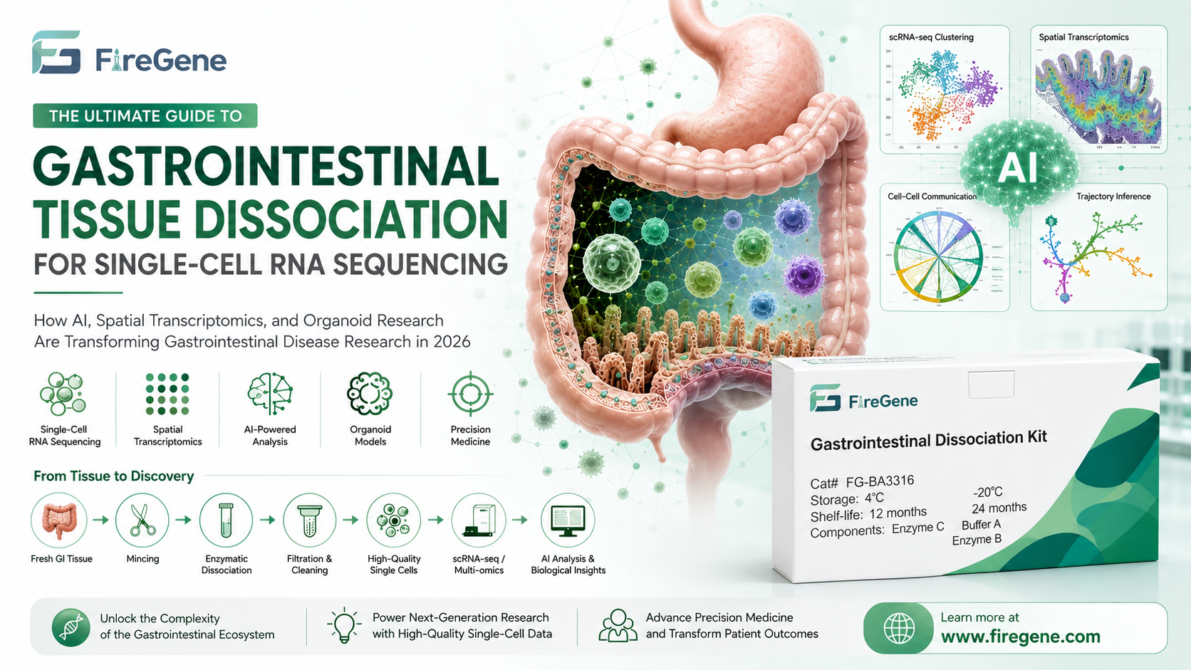
The Foundation of Every Successful Gastrointestinal Single-Cell Study Starts with Tissue Dissociation
Over the past decade, gastrointestinal (GI) research has undergone a remarkable transformation. Scientists are no longer limited to studying tissues as homogeneous structures. Instead, advances in single-cell RNA sequencing (scRNA-seq), spatial transcriptomics, multiplex imaging, organoid technology, and artificial intelligence (AI) now enable researchers to investigate the intestinal ecosystem at single-cell resolution.
Today, researchers can identify rare epithelial stem cells hidden within intestinal crypts, characterize immune cell dynamics during inflammatory bowel disease (IBD), map tumor microenvironments in colorectal cancer, and even predict therapeutic responses using AI-assisted computational models. These technologies are redefining our understanding of gastrointestinal biology and accelerating precision medicine.
However, despite these revolutionary analytical platforms, every successful single-cell experiment still depends on one fundamental step:
Obtaining a high-quality, viable single-cell suspension.
Regardless of whether the downstream workflow involves:
- 10x Genomics Chromium
- BD Rhapsody
- Drop-seq
- Smart-seq3
- Spatial transcriptomics
- Flow cytometry
- Organoid establishment
- CRISPR screening
- Multi-omics integration
the experiment can only be as good as the cells entering the workflow.
For gastrointestinal tissues, this initial preparation step is particularly challenging.
Unlike blood or cultured cells, gastrointestinal tissues contain:
- dense extracellular matrix (ECM)
- extensive mucus layers
- fragile epithelial structures
- tightly connected cell junctions
- abundant immune infiltrates
- diverse stromal populations
- region-specific architecture extending from stomach to colon
These characteristics make gut tissue one of the most difficult organs to dissociate into viable single cells without introducing technical bias.
Poor dissociation often results in:
- Low cell viability
- Selective loss of fragile epithelial populations
- Increased ambient RNA contamination
- Cell aggregation and doublets
- Stress-induced transcriptional artifacts
- Reduced sequencing efficiency
- Misleading biological interpretations
Consequently, tissue dissociation is no longer viewed as a routine sample preparation step—it is now recognized as a critical determinant of experimental success.
To address these challenges, specialized enzymatic workflows have largely replaced generic digestion protocols. Solutions such as the FireGene Gastrointestinal Dissociation Kit (FG-BA3316) are specifically optimized for gastrointestinal tissues, using a multi-step enzymatic strategy designed to generate high-quality, viable single-cell suspensions suitable for scRNA-seq, immune profiling, gut microbiome research, and organoid applications. The kit is formulated for mammalian GI tissues including stomach, small intestine, and colon, and is intended to preserve diverse epithelial and immune cell populations for downstream analyses.
In this comprehensive guide, we'll explore:
- Why gastrointestinal tissue is uniquely difficult to dissociate
- How dissociation quality directly influences single-cell sequencing results
- The growing role of AI in gastrointestinal single-cell biology
- Why spatial transcriptomics still depends on high-quality dissociated cells
- The connection between tissue dissociation and organoid success
- Best practices for maximizing cell recovery and viability
- Future trends shaping gastrointestinal disease research in 2026
Whether you're investigating inflammatory bowel disease, colorectal cancer, intestinal fibrosis, gastric cancer, or regenerative medicine, mastering gastrointestinal tissue dissociation is the essential first step toward generating reliable, biologically meaningful data.
Why Gastrointestinal Tissue Is One of the Most Difficult Organs to Dissociate
Researchers who routinely isolate cells from tissues such as the liver, spleen, or lymph nodes are often surprised by how much more challenging gastrointestinal tissue can be.
The reason lies in the extraordinary structural complexity of the gastrointestinal tract.
Unlike relatively uniform tissues, the GI tract must simultaneously perform digestion, nutrient absorption, immune surveillance, barrier protection, and continuous epithelial renewal. As a result, it contains one of the most diverse cellular ecosystems in the human body.
A typical intestinal biopsy includes dozens of distinct cell populations, including:
- intestinal stem cells
- enterocytes
- goblet cells
- Paneth cells
- enteroendocrine cells
- tuft cells
- fibroblasts
- endothelial cells
- smooth muscle cells
- macrophages
- dendritic cells
- T lymphocytes
- B lymphocytes
- innate lymphoid cells
- plasma cells
Each population differs dramatically in size, membrane composition, sensitivity to enzymatic digestion, and tolerance to mechanical stress.
This diversity makes it nearly impossible for a single harsh enzymatic protocol to preserve all cell types equally.
Dense Extracellular Matrix Limits Cell Release
The gastrointestinal extracellular matrix forms a highly organized scaffold composed of collagen, laminin, fibronectin, elastin, proteoglycans, and glycoproteins.
While this matrix maintains tissue integrity in vivo, it presents a major obstacle during cell isolation.
Insufficient digestion leaves many cells trapped within the matrix, leading to:
- low recovery rates,
- incomplete representation of stromal populations,
- poor reproducibility between samples.
Conversely, excessive enzymatic digestion can damage fragile epithelial cells, alter membrane proteins, and induce stress-response gene expression before sequencing even begins.
Achieving the correct balance between efficient ECM degradation and preservation of cell integrity is therefore essential.
The Protective Mucus Layer Creates Additional Challenges
Another unique feature of gastrointestinal tissue is its thick mucus coating.
Secreted primarily by goblet cells, mucus protects the intestinal epithelium against pathogens and mechanical injury. During tissue processing, however, mucus becomes problematic.
Excess mucus can:
- trap liberated cells,
- increase suspension viscosity,
- promote cell aggregation,
- interfere with filtration,
- reduce accurate cell counting,
- clog microfluidic devices used for scRNA-seq.
These issues significantly reduce sequencing efficiency and increase the likelihood of doublets.
Optimized gastrointestinal dissociation workflows therefore combine enzymatic digestion with carefully controlled washing and filtration steps to minimize mucus-related artifacts while preserving cell viability. FireGene's gastrointestinal workflow emphasizes sequential digestion and standardized filtration to improve recovery from complex, mucus-rich samples.
Why Cell Isolation Determines the Success of Single-Cell RNA Sequencing
Single-cell RNA sequencing has transformed biological research by allowing scientists to profile gene expression at the resolution of individual cells. Unlike bulk RNA sequencing, which averages signals across thousands or millions of cells, scRNA-seq captures cellular heterogeneity, enabling researchers to identify rare cell populations, reconstruct developmental trajectories, and uncover disease-specific molecular signatures.
However, one fundamental principle is often overlooked:
The quality of single-cell sequencing data is determined long before the sequencing run begins.
No sequencing platform, bioinformatics pipeline, or AI algorithm can recover biological information that was lost during tissue dissociation.
For gastrointestinal tissues, where epithelial cells are fragile and immune populations are highly diverse, obtaining a high-quality single-cell suspension is arguably the most important step in the entire workflow.
Cell Viability Directly Influences Sequencing Quality
The first metric researchers evaluate after tissue dissociation is cell viability.
High viability generally indicates that cells have maintained membrane integrity, intact RNA, and physiological gene expression profiles. Conversely, dead or dying cells release intracellular RNA into the surrounding solution, creating ambient RNA contamination.
Ambient RNA can lead to:
- False detection of genes in unrelated cell types
- Artificial inflation of gene counts
- Misclassification of cell identities
- Reduced clustering accuracy
- Increased background noise
For example, RNA released from dying epithelial cells may be mistakenly assigned to nearby immune cells, creating hybrid transcriptional profiles that do not exist biologically.
Modern computational tools such as SoupX, CellBender, and DecontX can partially reduce ambient RNA contamination, but they cannot completely restore the original biological signal. Prevention through optimized tissue dissociation remains far more effective than post hoc correction.
Mechanical Stress Can Alter the Transcriptome Before Sequencing
Another often underestimated factor is cellular stress induced during tissue processing.
Excessive mechanical disruption, prolonged enzymatic digestion, or inappropriate temperatures can activate immediate early response genes within minutes.
Genes such as:
- FOS
- JUN
- ATF3
- HSP family genes
may become highly expressed as a direct response to processing rather than disease biology.
If these stress signatures are not recognized, researchers may incorrectly interpret them as meaningful biological changes.
This issue is particularly important when studying inflammatory diseases such as Crohn's disease or ulcerative colitis, where stress-response pathways are already activated in vivo. Poor dissociation can therefore obscure genuine disease-associated transcriptional programs.
Fragile Cell Populations Are Easily Lost
Different gastrointestinal cell types tolerate enzymatic digestion differently.
Robust stromal cells often survive aggressive digestion, whereas fragile epithelial populations—including intestinal stem cells, goblet cells, and enteroendocrine cells—may be selectively damaged or lost.
This selective loss introduces sampling bias.
Instead of reflecting the true cellular composition of the tissue, the sequencing dataset becomes enriched for cells that simply survived the isolation process.
Such bias can affect downstream analyses, including:
- Differential abundance studies
- Cell–cell communication analysis
- Trajectory inference
- Pseudotime reconstruction
- Ligand–receptor interaction networks
Optimized tissue dissociation protocols aim to preserve both abundant and rare cell populations, ensuring that sequencing data accurately represent the original tissue architecture.
Doublets and Cell Aggregation Reduce Data Quality
Incomplete tissue digestion often leaves residual tissue fragments or cell aggregates.
These aggregates can enter droplet-based sequencing systems such as the 10x Genomics Chromium platform, where two or more cells may be encapsulated within the same droplet.
These doublets generate mixed transcriptomes that can appear as entirely new cell populations during downstream analysis.
Although computational algorithms such as DoubletFinder, Scrublet, and scDblFinder can identify many doublets, their performance decreases when doublet rates are high.
Careful tissue dissociation, filtration, and gentle handling remain the most effective strategies for minimizing these artifacts.
Every Bioinformatics Pipeline Depends on High-Quality Input
Modern scRNA-seq analysis pipelines—including Cell Ranger, Seurat, Scanpy, Bioconductor, and Harmony—assume that the input data faithfully represent the biology of the sample.
When tissue dissociation introduces excessive cell death, stress responses, or selective cell loss, these assumptions no longer hold.
As a result:
- Clusters become less distinct.
- Rare cell populations disappear.
- Batch effects increase.
- Cell annotations become less reliable.
- Downstream biological conclusions become less reproducible.
In other words:
Better computational analysis cannot compensate for poor sample preparation.
High-quality tissue dissociation is therefore the foundation upon which every successful single-cell study is built.
How AI Is Revolutionizing Gastrointestinal Single-Cell Research
Artificial intelligence has rapidly become one of the most influential technologies in biomedical research. While AI was once primarily associated with image recognition or clinical decision support, it is now reshaping nearly every stage of single-cell analysis—from automated cell annotation to drug target discovery.
In gastrointestinal research, where datasets routinely contain hundreds of thousands or even millions of cells, AI-driven tools have become indispensable for extracting meaningful biological insights.
From Manual Annotation to AI-Powered Cell Identification
Historically, identifying cell types in scRNA-seq datasets was a labor-intensive process.
Researchers manually compared marker gene expression against published literature, often spending days or weeks annotating a single dataset.
Today, AI-powered annotation platforms can classify cells within minutes.
Popular tools include:
- CellTypist
- scANVI
- scArches
- Azimuth
- GPT-assisted annotation workflows
- Large Cell Models (LCMs)
These systems compare gene expression profiles against large reference atlases and automatically assign probable cell identities with remarkable speed and consistency.
As reference atlases continue to expand, AI models are becoming increasingly capable of recognizing rare intestinal epithelial subsets, immune populations, stromal cells, and disease-associated transitional states.
AI Is Only as Good as the Data It Learns From
Despite these advances, AI models remain fundamentally dependent on data quality.
A machine learning algorithm cannot distinguish between genuine biological variation and technical artifacts if those artifacts dominate the training data.
Poor tissue dissociation introduces:
- Stress-response signatures
- Ambient RNA contamination
- Cell fragmentation
- Selective loss of fragile populations
- Artificial doublets
These issues can confuse AI models, leading to inaccurate cell classifications and misleading biological interpretations.
The familiar principle of machine learning applies perfectly to single-cell biology:
Garbage in, garbage out.
No matter how sophisticated the AI algorithm, its predictions can only be as reliable as the cells that entered the sequencing workflow.
AI Is Accelerating Drug Discovery
Beyond cell annotation, AI is transforming how researchers identify therapeutic targets.
By integrating single-cell transcriptomics with proteomics, genomics, spatial transcriptomics, and clinical outcomes, AI models can uncover molecular pathways associated with disease progression and treatment response.
In gastrointestinal diseases, AI-assisted analyses are helping researchers identify:
- Novel biomarkers for inflammatory bowel disease
- Immune checkpoints in colorectal cancer
- Fibrosis-associated stromal populations
- Predictors of immunotherapy response
- Regenerative pathways involved in mucosal healing
These discoveries are driving the development of precision medicine strategies tailored to individual patients.
However, the accuracy of these predictions still depends on high-quality biological input.
Every AI-driven insight ultimately begins with one essential requirement:
A representative, high-viability single-cell suspension generated through optimized tissue dissociation.
The Next Frontier: AI, Multi-Omics, and Foundation Models
The future of gastrointestinal research extends beyond transcriptomics alone.
Researchers are increasingly integrating:
- Single-cell RNA sequencing
- ATAC-seq
- Spatial transcriptomics
- Proteomics
- Metabolomics
- Digital pathology
- Clinical imaging
into unified multi-modal datasets.
Foundation AI models trained on millions of cells are expected to become the biological equivalent of large language models, capable of predicting disease progression, identifying therapeutic targets, and even simulating tissue responses to new drugs.
Yet regardless of how advanced these computational models become, they all share the same dependency:
Reliable biological data generated from high-quality tissue dissociation.
This principle will remain true as gastrointestinal research moves toward increasingly sophisticated AI-driven workflows.
Why Spatial Transcriptomics Still Depends on High-Quality Tissue Dissociation
Spatial transcriptomics has become one of the fastest-growing technologies in life science research. By preserving the spatial organization of tissues while measuring gene expression, it enables researchers to answer questions that conventional single-cell RNA sequencing cannot.
Instead of asking "Which genes are expressed?", spatial transcriptomics also reveals "Where are these genes expressed within the tissue?"
For gastrointestinal diseases, spatial information is particularly valuable because the intestinal microenvironment is highly organized.
Within only a few hundred micrometers, researchers can observe:
- Stem cells residing at the crypt base
- Transit-amplifying cells migrating upward
- Mature enterocytes lining the villi
- Immune cells infiltrating inflamed regions
- Fibroblast niches supporting epithelial regeneration
- Tumor cells interacting with stromal and immune compartments
Understanding these spatial relationships has become essential for studying inflammatory bowel disease (IBD), colorectal cancer (CRC), gastric cancer, intestinal fibrosis, and mucosal healing.
Can Spatial Transcriptomics Replace Single-Cell RNA Sequencing?
A common misconception is that spatial transcriptomics will eventually replace scRNA-seq. In reality, the two technologies are highly complementary.
Most spatial transcriptomics platforms profile gene expression from spots or pixels that often contain multiple cells. While newer technologies have improved resolution, many datasets still require single-cell reference atlases to accurately assign cell identities.
In other words, spatial transcriptomics depends on high-quality single-cell datasets to deconvolute mixed signals and interpret complex tissue architecture.
Without robust scRNA-seq references, researchers may struggle to distinguish closely related epithelial, stromal, or immune cell populations within spatial datasets.
This is why tissue dissociation remains indispensable, even in the era of spatial biology.
High-Quality Cell Isolation Supports Multi-Modal Analysis
Increasingly, researchers are combining:
- scRNA-seq
- Spatial transcriptomics
- Single-cell ATAC-seq
- Spatial proteomics
- Multiplex immunofluorescence
- Digital pathology
into integrated multi-modal workflows.
These approaches rely on consistent sample quality across different assays.
If the dissociated cell population is biased—for example, due to poor recovery of epithelial cells or excessive immune cell loss—the resulting reference atlas may compromise interpretation across all downstream datasets.
Optimized tissue dissociation therefore serves as the foundation for accurate integration of spatial and single-cell data.
Organoid Research Begins with Healthy Single Cells
Patient-derived organoids (PDOs) have rapidly become indispensable tools in gastrointestinal research.
These three-dimensional cultures retain many structural and functional characteristics of native tissues, allowing researchers to model disease progression, evaluate drug responses, and investigate regenerative mechanisms under controlled laboratory conditions.
Applications include:
- Colorectal cancer precision medicine
- Inflammatory bowel disease modeling
- Gastric cancer drug screening
- Intestinal stem cell biology
- Host–microbiome interactions
- Gene editing using CRISPR/Cas9
However, the success of organoid culture depends heavily on the quality of the starting cell population.
Why Tissue Dissociation Matters for Organoid Establishment
Unlike bulk tissue culture, organoids require viable progenitor and stem cell populations capable of self-renewal and differentiation.
Excessive enzymatic digestion or harsh mechanical processing can damage these delicate cells before they ever reach the culture plate.
Poor tissue dissociation may result in:
- Reduced organoid formation efficiency
- Slower growth rates
- Altered differentiation patterns
- Increased culture variability
- Premature organoid death
Conversely, gentle and optimized dissociation protocols help preserve epithelial stem cells while minimizing stress-induced damage.
This is particularly important when working with precious patient biopsy samples, where tissue availability is limited and experimental reproducibility is critical.
From Organoids to Precision Medicine
The integration of organoid technology with single-cell sequencing is transforming precision medicine.
Researchers can now:
- Establish patient-derived organoids.
- Perform scRNA-seq before and after treatment.
- Compare transcriptional changes at single-cell resolution.
- Predict therapeutic response.
- Identify mechanisms of drug resistance.
As artificial intelligence becomes increasingly integrated into these workflows, organoid-derived datasets are expected to play a central role in personalized treatment strategies for gastrointestinal diseases.
Best Practices for Successful Gastrointestinal Tissue Dissociation
Although every tissue sample presents unique challenges, several best practices consistently improve the quality of single-cell suspensions.
Process Fresh Tissue Whenever Possible
Cell viability declines rapidly after tissue collection.
Whenever feasible, begin tissue processing immediately after surgical resection or biopsy collection. If immediate processing is not possible, transport tissues in an appropriate preservation solution at low temperature while minimizing ischemic time.
Minimize Mechanical Damage
Mechanical disruption should be sufficient to expose tissue surfaces without excessively shearing cells.
Overly aggressive homogenization increases:
- Cell death
- RNA degradation
- Stress-response gene activation
- Doublet formation
Gentle mincing combined with optimized enzymatic digestion generally produces superior results.
Optimize Enzymatic Digestion
No single enzyme efficiently dissociates every gastrointestinal tissue.
Different tissue regions vary substantially in extracellular matrix composition, mucus content, and epithelial architecture.
Modern gastrointestinal dissociation workflows therefore rely on carefully balanced enzyme combinations rather than a single digestive enzyme.
The digestion time should be long enough to release intact cells while avoiding over-digestion that compromises membrane integrity.
Remove Cell Aggregates
Following enzymatic digestion, filtering the suspension through an appropriate cell strainer helps eliminate residual tissue fragments and large aggregates.
This step improves:
- Cell counting accuracy
- Microfluidic loading efficiency
- Sequencing performance
- Doublet reduction
Assess Cell Quality Before Sequencing
Before proceeding to library preparation, researchers should evaluate:
- Cell viability
- Cell concentration
- Aggregate frequency
- Debris levels
- Overall morphology
Investing a few additional minutes in quality control can prevent the loss of an entire sequencing experiment.
How the FireGene Gastrointestinal Dissociation Kit Supports Modern Single-Cell Research
As gastrointestinal single-cell technologies continue to evolve, researchers increasingly require tissue dissociation workflows that are reproducible, standardized, and compatible with diverse downstream applications.
The FireGene Gastrointestinal Dissociation Kit (FG-BA3316) was developed specifically to address the unique challenges associated with gastrointestinal tissues.
Rather than relying on generic enzymatic digestion protocols, the kit employs an optimized enzyme formulation designed for mammalian gastrointestinal tissues, helping researchers generate high-quality single-cell suspensions while preserving cellular diversity.
Key Features
✔ Optimized for gastrointestinal tissues, including stomach, small intestine, and colon
✔ Designed to preserve epithelial, immune, and stromal cell populations
✔ Supports high cell viability and recovery for downstream analyses
✔ Produces single-cell suspensions suitable for:
- Single-cell RNA sequencing (scRNA-seq)
- Single-cell ATAC-seq
- Spatial transcriptomics reference atlas construction
- Flow cytometry
- Cell sorting (FACS)
- Organoid establishment
- Primary cell culture
- Immune profiling
Typical Workflow
Fresh Gastrointestinal Tissue
↓
Mechanical Mincing
↓
FireGene Gastrointestinal Dissociation Kit (FG-BA3316)
↓
Gentle Enzymatic Digestion
↓
Filtration
↓
(Optional) Dead Cell Removal
↓
Cell Counting & Viability Assessment
↓
Single-Cell Sequencing / Flow Cytometry / Organoid Culture
This standardized workflow helps reduce experimental variability while supporting reproducible cell isolation across research projects.
Importantly, because the kit is compatible with multiple downstream applications, researchers can use a single optimized dissociation workflow to support diverse experimental pipelines, reducing the need for protocol redevelopment across different studies.
Future Trends: Where Gastrointestinal Single-Cell Research Is Heading Beyond 2026
The pace of innovation in gastrointestinal research continues to accelerate. Technologies that were once considered cutting-edge are rapidly becoming standard tools in both basic research and translational medicine. Looking ahead, several emerging trends are expected to reshape how scientists investigate gastrointestinal diseases and develop new therapies.
AI Will Become a Routine Laboratory Assistant
Artificial intelligence is evolving from a downstream analytical tool into an integrated research partner.
Future AI systems are expected to assist researchers throughout the experimental workflow by:
- Recommending optimized tissue dissociation parameters based on tissue type.
- Predicting cell viability before sequencing.
- Detecting technical artifacts during sample preparation.
- Automatically annotating newly discovered cell populations.
- Integrating transcriptomic, proteomic, and imaging datasets into unified biological models.
Rather than replacing laboratory expertise, AI will augment researchers' ability to interpret increasingly complex datasets.
Multi-Omics Integration Will Become the New Standard
Future gastrointestinal studies are unlikely to rely on transcriptomics alone.
Instead, researchers will routinely combine:
- Single-cell RNA sequencing
- Single-cell ATAC-seq
- Spatial transcriptomics
- Spatial proteomics
- Metabolomics
- Epigenomics
- Digital pathology
- Clinical imaging
These complementary datasets will provide a more comprehensive understanding of disease mechanisms than any single technology can achieve independently.
However, all of these approaches share one prerequisite:
High-quality biological samples.
Poor tissue dissociation cannot be corrected later through computational analysis or additional sequencing.
Digital Twins and Virtual Gastrointestinal Models
One of the most exciting developments is the concept of digital twins.
A digital twin is a computational model that represents an individual patient's gastrointestinal tissue using multi-modal biological data.
By integrating:
- Histology
- Imaging
- Single-cell sequencing
- Organoid drug-response data
- Clinical biomarkers
researchers may eventually simulate disease progression and predict therapeutic responses before treatment begins.
Although still in its early stages, digital twin technology has the potential to transform precision medicine for colorectal cancer, inflammatory bowel disease, and other gastrointestinal disorders.
Standardized Sample Preparation Will Become Increasingly Important
As international research collaborations continue to expand, reproducibility has become a major concern.
Large-scale initiatives increasingly require standardized tissue processing workflows to ensure that data generated across different laboratories remain comparable.
Optimized dissociation protocols help reduce technical variability and improve confidence in cross-study analyses.
For this reason, standardized gastrointestinal tissue dissociation is becoming an essential component of high-quality single-cell research rather than merely a laboratory technique.
Conclusion
Single-cell technologies have fundamentally changed our understanding of gastrointestinal biology.
Researchers can now investigate epithelial regeneration, immune responses, tumor evolution, stem cell dynamics, and host–microbiome interactions with unprecedented resolution.
Yet despite remarkable advances in sequencing technologies, artificial intelligence, spatial transcriptomics, and organoid biology, one principle remains unchanged:
Every successful single-cell experiment begins with high-quality tissue dissociation.
Obtaining a representative, viable, and unbiased single-cell suspension is essential for generating reliable sequencing data, constructing accurate spatial reference atlases, developing reproducible organoid models, and training robust AI algorithms.
As gastrointestinal research continues to embrace increasingly sophisticated multi-omics approaches, the importance of standardized sample preparation will only continue to grow.
The FireGene Gastrointestinal Dissociation Kit (FG-BA3316) was developed to support these evolving research needs by providing an optimized workflow for isolating high-quality single cells from mammalian gastrointestinal tissues. Whether your research focuses on inflammatory bowel disease, colorectal cancer, gastric disease, stem cell biology, or organoid development, a well-designed dissociation protocol lays the foundation for meaningful biological discovery.
Ultimately, breakthroughs in gastrointestinal medicine will not depend solely on faster sequencing platforms or more powerful AI models—they will depend on the quality of the biological samples from which every discovery begins.
Frequently Asked Questions (FAQ)
1. Why is gastrointestinal tissue difficult to dissociate?
Gastrointestinal tissue contains dense extracellular matrix, abundant mucus, tightly connected epithelial cells, diverse immune populations, and region-specific architecture. These characteristics make it one of the most challenging tissues for generating viable single-cell suspensions.
2. Why is tissue dissociation important for single-cell RNA sequencing?
Poor tissue dissociation can reduce cell viability, increase ambient RNA contamination, introduce stress-response artifacts, and selectively eliminate fragile cell populations. These issues directly affect sequencing quality and downstream biological interpretation.
3. What cell viability is generally recommended before scRNA-seq?
Although requirements vary by platform and study design, many laboratories aim for greater than 80–85% viability before library preparation to maximize data quality and minimize ambient RNA contamination.
4. Can over-digestion damage sequencing results?
Yes. Excessive enzymatic digestion may reduce membrane integrity, alter gene expression through cellular stress, and decrease recovery of sensitive epithelial and stem cell populations.
5. Can one dissociation protocol be used for all gastrointestinal tissues?
Not always. Stomach, small intestine, colon, and diseased tissues differ in extracellular matrix composition and cellular architecture. Optimized tissue-specific workflows generally provide more consistent results than universal protocols.
6. Does spatial transcriptomics eliminate the need for tissue dissociation?
No. Spatial transcriptomics and scRNA-seq are complementary technologies. High-quality single-cell datasets are still essential for creating reference atlases and accurately annotating spatial transcriptomic data.
7. Why are organoid studies sensitive to tissue dissociation quality?
Successful organoid cultures depend on viable epithelial stem and progenitor cells. Excessive mechanical or enzymatic damage during dissociation can significantly reduce organoid formation efficiency.
8. How does AI benefit gastrointestinal single-cell research?
AI accelerates cell annotation, identifies rare cell populations, predicts disease-associated pathways, integrates multi-omics datasets, and assists in therapeutic target discovery. Reliable AI models require high-quality biological input data.
9. What downstream applications require high-quality gastrointestinal single-cell suspensions?
High-quality suspensions support:
- Single-cell RNA sequencing
- Single-cell ATAC-seq
- Flow cytometry
- Cell sorting (FACS)
- Spatial transcriptomics reference generation
- Organoid culture
- Primary cell culture
- Immune profiling
- Multi-omics integration
10. What features should researchers consider when selecting a gastrointestinal tissue dissociation kit?
Researchers should evaluate tissue compatibility, cell viability, recovery efficiency, reproducibility, preservation of diverse cell populations, ease of use, and compatibility with downstream applications such as scRNA-seq and organoid culture.
FireGene Single-Cell Sample Prep
Looking for validated dissociation kits?
FireGene's tissue dissociation kits are optimized for specific organ types — brain, tumor, liver, GI, reproductive, and more. Validated for 10x Genomics Chromium and BD Rhapsody workflows.


