
Introduction
Organoid research has become an important technology for studying human development, tissue biology, disease mechanisms, and therapeutic responses. These three-dimensional, multicellular structures can reproduce selected features of their tissues of origin, making them valuable models for biomedical research.
However, successful organoid culture begins well before cells are embedded in an extracellular matrix or placed into an organoid growth medium. The quality of the starting cell population is one of the most important factors determining organoid performance.
Whether researchers are establishing intestinal crypts for gut organoid culture, isolating pancreatic islets for disease modeling, or characterizing intestinal epithelial populations by single-cell RNA sequencing (scRNA-seq), tissue dissociation directly affects cell recovery, viability, cell-type composition, and downstream experimental results.
This article explains the role of tissue dissociation in organoid research, the challenges associated with different tissue types, and how optimized dissociation workflows can support reliable organoid and single-cell applications.
Keywords: organoid research, organoid culture, tissue dissociation, organoid dissociation kit, single-cell RNA sequencing, scRNA-seq, intestinal organoids, tissue dissociation kit, cell isolation
1. What Is Organoid Research?
Organoids are three-dimensional cell cultures generated from stem cells, progenitor cells, or tissue-derived cells. Under appropriate culture conditions, these cells can self-organize into structures that reproduce certain characteristics of the original tissue.
Unlike conventional two-dimensional cell cultures, organoids can contain multiple cell types and display tissue-specific architecture and functions.
Organoid models are widely used in:
Developmental biology
Disease modeling
Cancer research
Drug discovery
Toxicology
Regenerative medicine
Personalized medicine
Single-cell analysis
For example, intestinal organoids can be used to study intestinal epithelial biology, host-pathogen interactions, inflammatory diseases, and drug responses. Pancreatic organoids and islet-derived models can support research into diabetes and pancreatic disorders, while liver, kidney, brain, and tumor organoids provide additional platforms for studying tissue-specific biology.
Despite the advantages of organoid technology, establishing a reproducible organoid model requires high-quality starting material. This makes tissue dissociation an essential part of the workflow.
2. Why Tissue Dissociation Matters in Organoid Research
Before organoids can be established, researchers often need to release cells, crypts, islets, or other tissue structures from their native environment.
Biological tissues are complex structures composed of cells surrounded by extracellular matrix (ECM), cell-cell junctions, and other structural components. Enzymatic and mechanical dissociation methods are commonly used to break down these interactions and generate the desired starting material.
The dissociation process can influence several critical parameters.

Cell Viability
Excessive mechanical force or prolonged enzymatic exposure can damage cells and reduce viability. Low-viability starting populations may result in poor organoid formation or inconsistent culture performance.
Cell Recovery
An inefficient dissociation procedure may leave valuable cells trapped within tissue fragments. Improving recovery can increase the number of viable cells available for organoid establishment.
Cell-Type Representation
Different cell populations may have different sensitivities to enzymatic treatment. Over-digestion may selectively damage certain populations, potentially changing the cellular composition of the final suspension.
Cellular State
Processing conditions can also affect cellular stress responses and gene expression profiles. This is particularly important when cells will subsequently be analyzed using scRNA-seq or other transcriptomic technologies.
Therefore, the goal of tissue dissociation is not simply to produce a single-cell suspension. The objective is to obtain a high-quality, representative cell population while minimizing unnecessary cellular damage.
3. Tissue Dissociation for Different Organoid Models
Different tissues have different structural characteristics, meaning that dissociation requirements can vary considerably.
Intestinal Organoids
Intestinal organoid research frequently involves the isolation of intestinal crypts or epithelial cells. The intestinal epithelium contains multiple specialized populations, including stem cells, absorptive cells, secretory cells, and other epithelial subtypes.
An optimized dissociation workflow can help researchers recover intact crypt structures or viable epithelial cells while minimizing excessive tissue damage.
These preparations can subsequently be used for:
Gut organoid establishment
Intestinal stem cell research
Epithelial differentiation studies
Disease modeling
Drug screening
scRNA-seq
Pancreatic Organoids and Islet Research
The pancreas contains multiple cell populations embedded within a complex tissue structure. Efficient tissue processing can be important for recovering pancreatic cells or islet structures for downstream research.
Researchers may use these preparations to investigate pancreatic development, β-cell biology, diabetes, and drug responses.
Tumor Organoids
Tumor tissues are highly heterogeneous and can contain cancer cells, stromal cells, immune cells, endothelial cells, and other populations.
Dissociation quality can therefore influence which populations are recovered for tumor organoid establishment or single-cell profiling. Gentle and appropriately optimized processing can help preserve cellular diversity and improve the biological relevance of downstream analyses.
Brain and Other Complex Tissues
Neural tissues and other dense organs can present additional dissociation challenges because of their extracellular matrix, cell-cell interactions, and sensitivity to processing conditions.
For these applications, tissue-specific dissociation strategies can help researchers obtain viable cells or nuclei for organoid research and single-cell analysis.
4. Tissue Dissociation and Single-Cell RNA Sequencing
Organoid research is increasingly combined with single-cell RNA sequencing (scRNA-seq) to investigate cellular heterogeneity at high resolution.
A typical workflow may include:
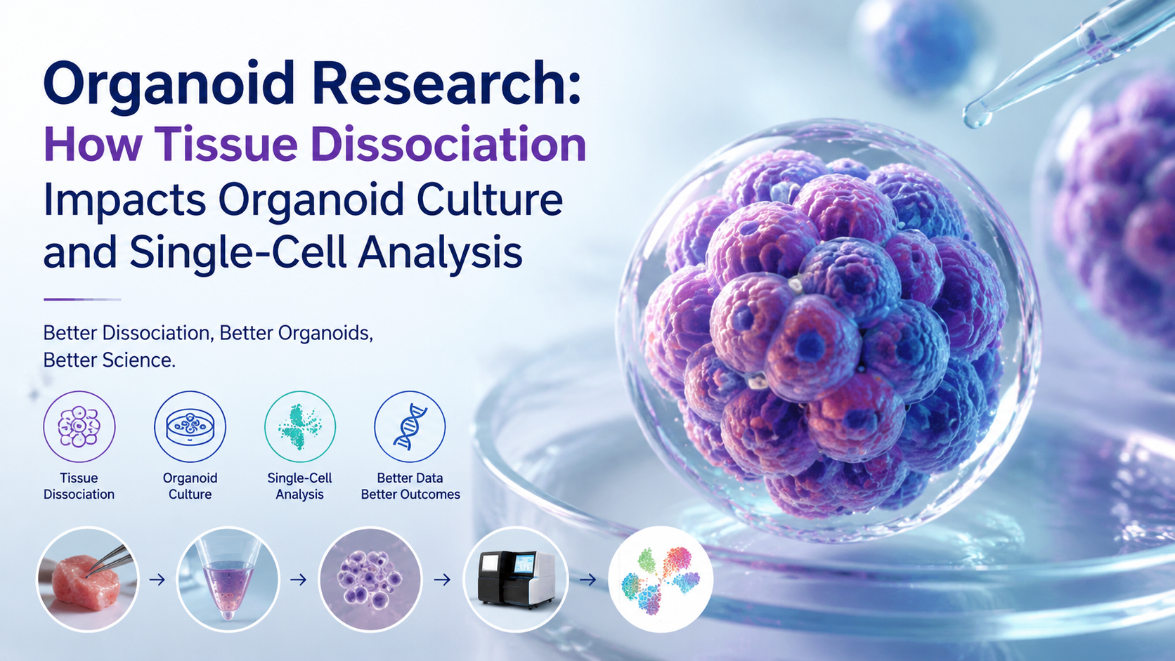
Tissue → Dissociation → Cell suspension → Quality control → Library preparation → scRNA-seq → Bioinformatics analysis
The dissociation step occurs at the beginning of this workflow, but its effects can extend throughout the entire experiment.
A poor-quality cell suspension may contain excessive cell debris, dead cells, aggregates, or underrepresented cell populations. These issues can reduce the quality of sequencing libraries and complicate downstream data analysis.
For organoid studies, scRNA-seq can help researchers investigate:
Cell-type composition
Cellular differentiation
Stem and progenitor populations
Disease-associated cell states
Cell-cell communication
Developmental trajectories
Treatment responses
Gene expression profiles
Importantly, researchers should consider that dissociation itself can introduce cellular stress. Optimizing tissue handling, digestion conditions, temperature, processing time, and downstream recovery can help minimize unwanted technical effects.
5. How to Optimize Tissue Dissociation for Organoid Research
There is no universal dissociation protocol that works equally well for every tissue. Optimization should take into account the tissue type, desired cell population, downstream application, and required degree of dissociation.
5.1 Select a Tissue-Specific Dissociation Strategy
The first step is choosing a dissociation method appropriate for the target tissue. Different tissues may require different combinations of enzymatic digestion and mechanical disruption.
5.2 Balance Digestion Efficiency and Cell Viability
More aggressive digestion does not necessarily produce better results. Excessive digestion may reduce viability or damage important surface markers.
The objective is to achieve sufficient tissue disruption while maintaining the biological quality of the recovered cells.
5.3 Minimize Processing Stress
Tissue should generally be processed efficiently and consistently. Unnecessary delays, excessive mechanical manipulation, or prolonged exposure to digestive enzymes can negatively affect cell quality.
5.4 Remove Debris and Aggregates
Following dissociation, filtration and appropriate cell-cleanup procedures can help improve suspension quality. Removing large debris and unwanted aggregates can facilitate downstream cell counting, organoid establishment, and single-cell sequencing.
5.5 Evaluate Cell Quality Before Culture or Sequencing
Important quality-control parameters may include:
Total cell yield
Cell viability
Cell concentration
Aggregation
Debris levels
Morphology
Cell-type representation
For scRNA-seq applications, maintaining a clean and viable cell suspension is particularly important because poor starting material can affect sequencing efficiency and data interpretation.
6. Advantages of Optimized Dissociation Kits
Using a validated tissue dissociation kit can simplify experimental workflows and improve consistency compared with developing every digestion condition from scratch.
A well-designed dissociation kit may provide optimized enzyme combinations and processing conditions for specific tissues or applications.
Potential advantages include:
Consistent Cell Recovery
Standardized reagents and protocols can reduce variation between experiments.
Improved Cell Viability
Optimized digestion conditions can help minimize unnecessary cellular damage.
Better Reproducibility
Reproducible tissue processing is particularly valuable for longitudinal studies, disease models, and high-throughput experiments.
Compatibility with Downstream Applications
A suitable dissociation workflow can support multiple applications, including organoid culture, flow cytometry, cell sorting, and scRNA-seq.
For researchers working with multiple tissue types, tissue-specific dissociation solutions can also reduce the need for extensive protocol optimization.
7. Organoid Research Applications
High-quality tissue dissociation supports a broad range of organoid research applications.
Disease Modeling
Organoids can reproduce selected disease-associated phenotypes and provide experimental models for investigating disease mechanisms.
Drug Discovery
Researchers can use organoids to evaluate compound activity, toxicity, and treatment responses in more physiologically relevant three-dimensional systems.
Personalized Medicine
Patient-derived organoids may provide an experimental platform for evaluating individual responses to therapeutic interventions.
Regenerative Medicine
Organoid systems can help researchers investigate stem cell differentiation, tissue development, and regeneration.
Single-Cell Profiling
Combining organoid models with scRNA-seq enables detailed characterization of cellular populations and changes during development, disease progression, or treatment.
Conclusion
The success of organoid research depends heavily on the quality of the cells or tissue structures used to initiate the culture. Tissue dissociation is therefore more than a sample-preparation step—it is a critical determinant of downstream experimental quality.
An optimized dissociation workflow can improve cell recovery, maintain viability, preserve representative cell populations, and provide high-quality starting material for organoid culture and single-cell analysis.
As organoid technology continues to expand across intestinal, pancreatic, tumor, neural, hepatic, and other research fields, reliable tissue dissociation solutions will remain an important component of reproducible workflows.
For researchers establishing organoid cultures or integrating organoids with scRNA-seq, selecting an appropriate tissue dissociation kit and optimizing the cell preparation process can help create more consistent, biologically relevant experimental models.


