
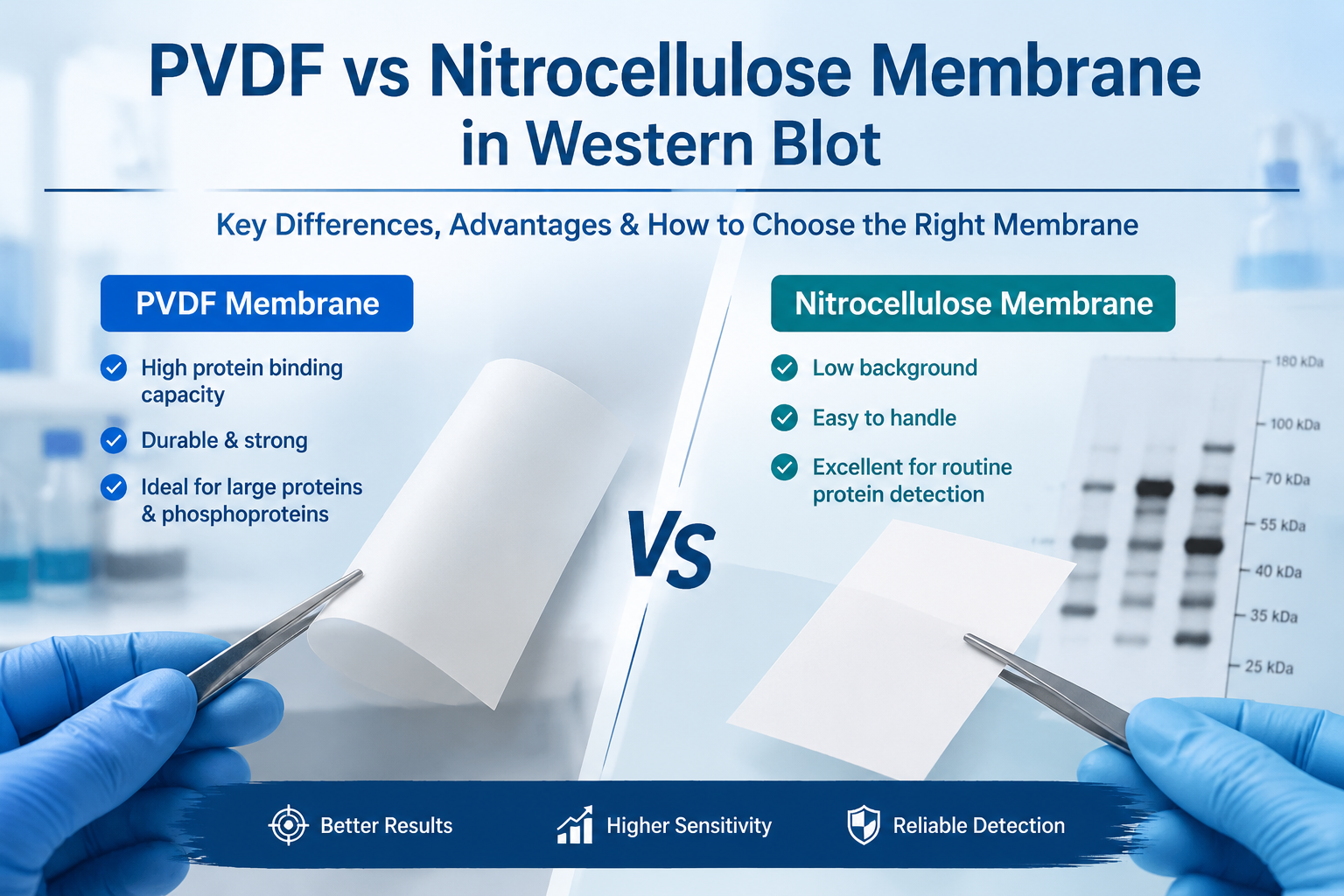
Introduction
Western blot (WB) is one of the most widely used techniques in molecular biology for detecting and analyzing specific proteins in complex biological samples. A successful Western blot experiment depends on multiple factors, including protein extraction, gel electrophoresis, transfer efficiency, antibody quality, and detection methods. Among these factors, the selection of the right transfer membrane plays a critical role in achieving reliable and reproducible results.
During the Western blot transfer step, proteins separated by SDS-PAGE are transferred from the polyacrylamide gel onto a membrane where they can be detected by specific antibodies. The two most commonly used transfer membranes are PVDF membrane (polyvinylidene fluoride membrane) and nitrocellulose membrane (NC membrane).
Although both membranes are widely used for Western blotting, they differ in protein-binding capacity, mechanical strength, background performance, and suitability for different protein targets. Understanding the advantages and limitations of PVDF and NC membranes can help researchers select the most appropriate membrane for their experiments.
What Is a Western Blot Transfer Membrane?
A Western blot transfer membrane is a solid support material used to immobilize proteins after electrophoretic separation. During the transfer process, proteins migrate from the gel onto the membrane under an electric field. Once immobilized, proteins can be detected through antibody-based methods, including:
· Primary antibody binding
· Secondary antibody recognition
· Chemiluminescent or fluorescent signal detection
The quality of the transfer membrane directly affects:
· Protein retention
· Signal intensity
· Background noise
· Detection sensitivity
· Reproducibility of experimental results
The two major membrane materials used in WB are:
1. PVDF membrane
2. Nitrocellulose membrane
PVDF Membrane in Western Blot
What Is PVDF Membrane?
PVDF membrane is a synthetic fluoropolymer membrane widely used in protein analysis. It has strong hydrophobic properties that allow efficient binding of proteins through hydrophobic interactions.
PVDF membranes are available in different pore sizes, commonly:
· 0.45 μm for most Western blot applications
· 0.2 μm for small proteins and high-resolution detection
Because of its excellent durability and high protein-binding capacity, PVDF has become a preferred choice for many advanced Western blot applications.
Advantages of PVDF Membrane
1. High Protein Binding Capacity
One of the major advantages of PVDF membrane is its high protein-binding ability.
Compared with nitrocellulose membranes, PVDF can immobilize larger amounts of protein, making it particularly useful for:
· Low-abundance proteins
· Weakly expressed targets
· Multiple detection procedures
The strong protein retention capability helps improve detection sensitivity, especially when protein levels are limited.
2. Excellent Mechanical Strength
PVDF membranes are highly durable and resistant to tearing. This makes them suitable for:
· Long experimental procedures
· Multiple antibody stripping and reprobing
· Long-term membrane storage
For researchers who need to detect multiple proteins from the same blot, PVDF provides better flexibility and stability.
3. Ideal for Large Molecular Weight Proteins
Large proteins often transfer inefficiently because of their size. PVDF membranes provide stronger protein retention and are commonly recommended for proteins above 100 kDa.
Examples include:
· mTOR (~289 kDa)
· Myosin proteins
· Large receptor proteins
· Cytoskeletal proteins
Using PVDF can help reduce protein loss during washing and antibody incubation steps.
4. Suitable for Phosphorylated Proteins
Phosphorylation-specific Western blotting requires careful preservation of protein modifications. PVDF membranes are commonly used for phosphoprotein detection because of their strong protein-binding characteristics.
Common applications include:
· Phospho-AKT detection
· Phospho-ERK analysis
· Phospho-mTOR studies
· Cell signaling pathway research 
Limitations of PVDF Membrane
Despite its advantages, PVDF also has some limitations.
1. Requires Methanol Activation
Because PVDF is naturally hydrophobic, it must be activated before use.
A typical preparation process includes:
1. Wetting the membrane with 100% methanol
2. Rinsing with distilled water
3. Equilibrating in transfer buffer
Without proper activation, protein transfer efficiency may decrease.
2. Higher Cost
PVDF membranes are generally more expensive than nitrocellulose membranes. For routine Western blot experiments involving abundant proteins, NC membranes may provide a more economical option.
Nitrocellulose Membrane in Western Blot
What Is Nitrocellulose Membrane?
Nitrocellulose membrane is one of the earliest membrane materials developed for protein immobilization. It has been widely used in Western blotting due to its excellent protein-binding properties and low background signal.
NC membranes bind proteins mainly through hydrophobic interactions and electrostatic forces.
Advantages of Nitrocellulose Membrane
1. Low Background Signal
A major advantage of nitrocellulose membranes is their clean background performance.
NC membranes generally provide:
· Low nonspecific binding
· Clear antibody signals
· Good signal-to-noise ratios
This makes them suitable for routine protein detection and quantitative Western blot analysis.
2. Easy Handling
Unlike PVDF membranes, nitrocellulose membranes do not require methanol activation.
The basic preparation process includes:
· Equilibration in transfer buffer
· Direct placement into the transfer system
This makes NC membranes convenient, especially for beginners or high-throughput laboratories.
3. Excellent for Routine Protein Detection
NC membranes perform very well for commonly analyzed proteins in the range of approximately 20–100 kDa.
Typical targets include:
· GAPDH (~36 kDa)
· β-actin (~42 kDa)
· Tubulin (~55 kDa)
For standard protein expression analysis, NC membranes remain a reliable and widely used option.
Limitations of Nitrocellulose Membrane
1. Lower Mechanical Strength
Nitrocellulose membranes are more fragile than PVDF membranes.
They can:
· Tear easily
· Crack when dry
· Require careful handling
This makes them less suitable for repeated stripping and reuse.
2. Lower Protein Binding Capacity
Although NC membranes bind proteins efficiently, their binding capacity is lower than PVDF membranes.
For:
· Very low-expression proteins
· Large proteins
· Multiple detection experiments
PVDF may provide better results.
PVDF vs Nitrocellulose Membrane: Key Differences
|
Feature |
PVDF Membrane |
Nitrocellulose Membrane |
|
Protein binding capacity |
Higher |
Moderate |
|
Mechanical strength |
Excellent |
Lower |
|
Background signal |
Low |
Very low |
|
Protein retention |
Strong |
Good |
|
Large protein detection |
Excellent |
Moderate |
|
Small protein detection |
Requires optimization |
Good |
|
Methanol activation |
Required |
Not required |
|
Stripping and reprobing |
Recommended |
Limited |
|
Cost |
Higher |
Lower |
|
Common applications |
Advanced WB, phosphoproteins, low abundance targets |
Routine WB analysis |
How to Choose Between PVDF and Nitrocellulose Membrane?
The choice between PVDF and NC membrane depends mainly on the experimental goal.
Choose PVDF Membrane When:
· Detecting low-abundance proteins
· Working with proteins larger than 100 kDa
· Studying phosphorylation pathways
· Performing multiple antibody detections
· Preparing publication-quality Western blot data
Choose Nitrocellulose Membrane When:
· Performing routine Western blot experiments
· Detecting highly expressed proteins
· Looking for low background signals
· Working with limited budgets
· Conducting simple protein expression analysis
Tips for Improving Western Blot Transfer Efficiency
Regardless of membrane selection, several factors influence transfer performance:
1. Optimize Transfer Conditions
Transfer parameters depend on:
· Protein molecular weight
· Gel percentage
· Transfer method
· Membrane type
Large proteins often require longer transfer times, while small proteins require shorter transfer conditions.
2. Select the Appropriate Membrane Pore Size
Common recommendations:
· 0.45 μm membrane: Most proteins
· 0.2 μm membrane: Small proteins (<20 kDa)
Selecting the correct pore size helps prevent protein loss and improves detection sensitivity.
3. Avoid Excessive Transfer
Over-transfer can cause proteins to pass through the membrane, especially with small molecular weight proteins.
Optimizing transfer time is essential for obtaining accurate results.
Conclusion
Both PVDF and nitrocellulose membranes are valuable tools for Western blot analysis. The best choice depends on the protein target, experimental requirements, and detection strategy.
PVDF membranes provide superior protein-binding capacity, durability, and performance for challenging applications such as large proteins, low-expression targets, and phosphoprotein studies. Nitrocellulose membranes offer excellent background performance, easy handling, and cost efficiency for routine Western blot experiments.
By understanding the differences between PVDF and NC membranes, researchers can select the most suitable transfer membrane to improve Western blot sensitivity, accuracy, and reproducibility.


