
Description
Overview
FireGene Human Brain Dissociation Kit is a high-efficiency enzymatic system designed to dissociate human brain tissue into high-viability single-cell suspensions. Ideal for neurological research and single-cell sequencing, this kit enables accurate profiling of brain cell populations involved in health and disease.
Background Information
-
Driven by Clinical and Scientific Research Needs:
- Single-cell sequencing of human brain tissue is essential for understanding complex neural circuits and brain disorders.
- Traditional dissociation methods struggle with preserving diverse brain cell types such as neurons and glia.
- This kit enables:
- High-resolution analysis of cellular heterogeneity in both healthy and diseased brain tissues.
- Identification of biomarkers and therapeutic targets for conditions like neurodegeneration, epilepsy, and mental health disorders.
- Advancement of precision neurology through better cellular insight.
-
Background of Technological Development:
- Older dissociation methods often lead to low cell yields and viability.
- FireGene’s solution:
- Uses a synergistic enzymatic blend optimized for fragile neural tissue.
- Features carefully calibrated enzyme types, concentrations, and temperature conditions.
- Maximizes viable cell recovery for reliable, reproducible results in downstream assays.
Detection Principle
- Utilizes a stepwise enzymatic digestion method:
- Brain samples are cut into small pieces to enhance enzyme penetration.
- Enzymes are added sequentially under optimal incubation conditions.
- Extracellular matrix and intercellular junctions are enzymatically broken down.
- Result:
- A clean, viable single-cell suspension suitable for scRNA-seq, brain atlas mapping, flow cytometry, or culture.
Specifications
| Applications | Single-cell sequencing, cell culture or other cell-related detections |
| Compatible Sample Types | Human brain tissue |
| Supported Instruments | Water bath, horizontal centrifuge, cell counter |
| Storage | -20 °C / 4 °C |
| Shelf-life | 24 months at -20 °C 12 months at 4 °C |
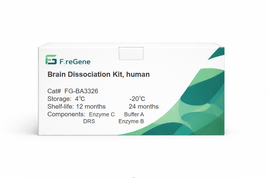
Kit Components
10 Reactions
| Component | 10 Reactions/Kit | Storage | Shelf-life |
|---|---|---|---|
| Buffer A | 1*21.6 mL | -20 °C | 24 months |
| Enzyme B | 1*8 mL | -20 °C | 24 months |
| Enzyme C | 1*100 μL | 4 °C | 12 months |
| DRS (Human Brain Cell Debris Removal Buffer) | 1*10 mL | 4 °C | 12 months |
50 Reactions
| Component | 50 Reactions/Kit | Storage | Shelf-life |
|---|---|---|---|
| Buffer A | 5*21.6 mL | -20 °C | 24 months |
| Enzyme B | 5*8 mL | -20 °C | 24 months |
| Enzyme C | 5*100 μL | 4 °C | 12 months |
| DRS (Human Brain Cell Debris Removal Buffer) | 5*10 mL | 4 °C | 12 months |
Product FAQ
1. Q: When dissociating human brain tissue, flocculent precipitates appear immediately after adding Enzyme C (10μL/reaction). Will this affect the enzymolysis effect? How to handle the precipitation issue?
A: Flocculent precipitates are mostly caused by a temporary reaction between Enzyme C (enzyme preparation) and ionic components in Buffer A. If the precipitates dissolve on their own within 10 minutes, the enzymolysis effect will not be affected. If the precipitates persist, reconfigure the reaction system: ① First, mix 10μL Enzyme C with 800μL Enzyme B uniformly to form an "enzyme mixture"; ② Slowly add the mixture dropwise into Buffer A containing the tissue, and invert to mix while adding. This avoids precipitation caused by local high concentration and ensures stable enzymolysis efficiency.
2. Q: After adding DRS (Debris Removal Solution) in Step 11, the cell suspension is pipetted 10 times. What impact will excessive pipetting force or insufficient pipetting times have on cell separation? How to define the standard for "gentle pipetting"?
A: Excessive pipetting force: Damages the cell membrane of fragile cells such as neurons and glial cells, reducing cell viability by 30%-40%, with a large number of cell debris visible under the microscope. Insufficient pipetting times (<8 times): DRS cannot fully contact the cell suspension, so debris cannot float up effectively during subsequent centrifugation and stratification, reducing cell purity by 50%. Standard for "gentle pipetting": Use a 5mL pipette, insert the pipette tip slowly 1cm below the liquid surface when aspirating, control the aspiration time to 2 seconds, and push the liquid out slowly along the tube wall when dispensing. Avoid generating air bubbles and ensure stable, non-impact liquid flow.
3. Q: The centrifugation parameters in Step 13 are "4℃, 3000×g, 20 minutes, medium acceleration/deceleration". If the laboratory's horizontal centrifuge does not have the "medium acceleration/deceleration" function and only has "fast" or "slow" options, how to adjust the operation?
A: If medium acceleration/deceleration is unavailable, prioritize "slow acceleration/deceleration" (acceleration time ≥5 minutes, deceleration time ≥8 minutes) and extend the centrifugation time to 25 minutes: ① Slow acceleration prevents liquid disturbance due to sudden increase in centrifugal force, ensuring stable stratification of DRS and cells; ② Slow deceleration prevents bottom cells from being scattered by inertia, reducing mixing with the debris layer. If only fast acceleration/deceleration is available, let the centrifuge tube stand for 2-5 minutes after centrifugation before taking it out to stabilize the stratification interface. This reduces the probability of cell-debris mixing by 30%, but the cell yield will still be approximately 15% lower than that with medium acceleration/deceleration.
4. Q: When dissociating aged human brain tissue (refrigerated for more than 6 hours after sampling), the cell viability is only 30%, much lower than the 70% of fresh tissue. How to optimize the operation to improve the cell viability of aged tissue?
A: The low viability of aged tissue requires optimization from two aspects: "reducing cell damage" and "enhancing cell protection": ① Immediately soak the tissue in PBS containing 10% FBS (5% for regular use) after sampling, and transport it under refrigeration at 4℃ to avoid tissue dehydration; ② Reduce the amount of Enzyme B to 600μL (800μL for regular use) during enzymolysis, and shorten the enzymolysis time to 15-20 minutes to reduce enzyme-induced damage to fragile cells; ③ Replace regular PBS with PBS containing 1% BSA in the washing step to enhance cell membrane protection. These measures can increase the cell viability of aged tissue to over 50%.
5. Q: After adding red blood cell lysis buffer (FG-BA3311) in Step 15, the mixture is incubated on ice for 5 minutes. What impact will prolonged incubation (e.g., 10 minutes) or insufficient incubation (e.g., 2 minutes) have on human brain cells (e.g., neurons)?
A: Prolonged incubation: Red blood cell lysis buffer has mild toxicity to neurons; 10-minute incubation reduces neuron viability by 25%-30%, and glial cells tend to shrink. Insufficient incubation: Red blood cells are not fully lysed, with residual red blood cells accounting for over 20%, which interferes with cell capture in subsequent single-cell sequencing (non-specific adsorption to the sequencing chip). The optimal operation is to observe under a microscope every 2 minutes and terminate the incubation immediately when red blood cells become transparent (approximately 4-5 minutes). This balances the effect of red blood cell removal and cell viability protection.
6. Q: Step 20 mentions "if obvious cell clumping occurs, filter with a 20μm cell sieve". However, after filtration, a large number of neurons (with a diameter of approximately 8-12μm) are retained. What is the reason, and how to adjust the filtration operation?
A: Neurons are retained mostly because they form clumps >20μm by entangling with glial cells and debris, rather than single neurons being retained. Adjustment methods: ① Before filtration, gently pipette the cell suspension 15 times with a 1mL low-adhesion pipette tip (avoid violence) to disperse small clumps <20μm; ② Pre-wet the 20μm cell sieve with PBS containing 5% FBS to reduce cell adsorption to the sieve; ③ Gently push the liquid with a 5mL syringe during filtration to avoid clump compression caused by sieve clogging. These measures can reduce the neuron retention rate from 40% to below 10%.
7. Q: After opening Buffer A in the kit, it is stored at 4℃ for 1 month. When used, slight turbidity is found in the solution. Is it still usable? How to quickly determine if the turbid Buffer A is invalid?
A: Slight turbidity may be caused by the low-temperature precipitation of components in Buffer A. Its validity needs to be verified first: ① Take 100μL of turbid Buffer A and heat it in a 37℃ water bath for 10 minutes. If the turbidity disappears, it indicates normal precipitation; the buffer can be used after shaking uniformly. ② If the turbidity persists after heating or flocculent precipitates appear, Buffer A is contaminated or deteriorated and cannot be used. To avoid turbidity after opening, aliquot Buffer A into 2mL/tube (one tube for one experiment) after opening, store it frozen at -20℃, and thaw and mix well before each use. This extends the validity period to 3 months.
8. Q: A 70μm cell sieve is used for filtration in Steps 5 and 6. If the sieve is severely clogged, the filtration speed is extremely slow, and a large amount of white flocculent material (suspected to be nerve fibers) remains on the sieve, how to pretreat to reduce nerve fiber residue?
A: The white flocculent material is nerve fibers. A "pre-digestion" step needs to be added before enzymolysis: ① After mincing the tissue, soak it in 2160μL Buffer A for 10 minutes, and gently shake every 5 minutes; ② Add 50μL Enzyme B (1/16 of the regular 800μL), incubate at 37℃ for 5 minutes to preliminarily degrade nerve fibers; ③ Add the remaining Enzyme B and Enzyme C according to the regular procedure. This reduces nerve fiber formation by 60%, avoids sieve clogging, and increases the filtration speed by 40%.
9. Q: After quality control, it is found that glial cells account for 90% of the cell suspension, while neurons only account for 10%—much lower than the normal neuron proportion in brain tissue (approximately 30%). What causes neuron loss, and how to optimize the collection method?
A: Neuron loss is mostly due to "weak neuron sedimentation capacity" in centrifugation and filtration steps: ① In Step 7, increase the centrifugation speed from 300×g to 350×g and extend the centrifugation time to 8 minutes to enhance neuron precipitation; ② Before filtration in Step 20, if there is no obvious cell clumping, skip the 20μm cell sieve filtration (neurons are easily adsorbed by the sieve); ③ When aspirating the supernatant in Step 14, retain 100μL of supernatant to mix with the cell pellet to avoid the loss of small-volume neurons with the supernatant. These measures can increase the neuron proportion to approximately 25%.
Files Download Links