
Introduction
Lung organoids have become valuable experimental models for investigating pathological changes in pulmonary diseases. Compared with conventional two-dimensional cell culture, three-dimensional lung organoid systems can better reproduce important characteristics of pulmonary epithelial organization, cell–cell interactions, tissue remodeling, and disease-associated phenotypes.
For organoid models established directly from pathological lung tissue, however, one of the first technical challenges occurs before the cells are embedded in an extracellular matrix or exposed to organoid culture medium: the original lung tissue must be dissociated efficiently while maintaining the viability and biological characteristics of organoid-forming cells.
This requirement becomes particularly important when working with diseased lung specimens. Fibrosis, mucus accumulation, inflammatory infiltration, altered extracellular matrix composition, and structural remodeling can make pathological lung tissue more difficult to dissociate than normal tissue.
The FireGene Lung Dissociation Kit provides a lung-specific enzymatic approach for converting lung tissue into viable cellular preparations suitable for subsequent cell culture. In a pathological lung organoid workflow, the kit can therefore serve as the critical tissue-preparation step connecting the original disease specimen with downstream three-dimensional organoid culture.
FireGene describes the kit as using an optimized enzyme combination adapted to lung extracellular matrix composition, with controlled digestion conditions intended to improve cell recovery while reducing excessive cellular stress. The resulting cells can subsequently enter cell-culture workflows.
Why Tissue Dissociation Matters in Pathological Lung Organoid Establishment
Establishing an organoid from primary lung tissue involves more than simply placing a tissue fragment into Matrigel.
The organoid-forming epithelial cells must first be released from the complex structural environment of the lung.
Normal lung tissue already contains a highly organized architecture consisting of alveolar structures, airways, connective tissue, vascular networks, basement membranes, and extracellular matrix proteins. Pathological processes can make this architecture considerably more difficult to process.
For example, fibrotic lung tissue may contain extensive extracellular matrix deposition and remodeled collagen-rich regions. COPD-associated tissue may contain increased mucus and changes in airway structure. Chronically inflamed tissue may contain substantial inflammatory infiltration, cellular debris, and damaged tissue regions.
These alterations directly affect how efficiently cells can be recovered.
Recent work establishing three-dimensional organoids from fibrotic human lung tissue illustrates the importance of this initial dissociation step. Fibrotic peripheral lung tissue was mechanically minced and enzymatically digested before alveolar epithelial cells were isolated and subsequently embedded in Matrigel for organoid culture.
This means that tissue dissociation is not merely a preliminary handling procedure. It can influence the quality of the starting cell population from which the pathological organoid model is ultimately established.
Role of the FireGene Lung Dissociation Kit
The FireGene Lung Dissociation Kit is designed specifically for enzymatic dissociation of lung tissue.
According to the manufacturer's technical information, the workflow involves reducing lung tissue into small fragments and exposing the tissue to a synergistic enzymatic digestion system. The enzymes disrupt extracellular matrix components and cell–matrix interactions, allowing viable pulmonary cells to be released from the original tissue structure.
Within a pathological lung organoid workflow, its role can be summarized as:
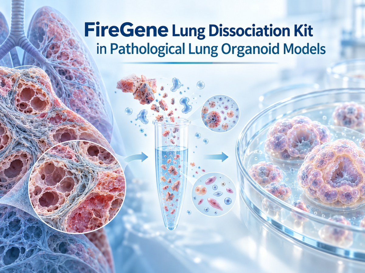
Pathological Lung Tissue → Controlled Tissue Dissociation → Viable Pulmonary Cell Preparation → Organoid-Forming Cell Population → ECM Embedding → Pathological Lung Organoid
The FireGene kit therefore operates at the transition between the original pathological specimen and the organoid culture system.
It does not itself generate the organoid. Instead, it helps provide the cellular starting material required for successful organoid establishment.
This distinction is important because organoid formation subsequently depends on additional factors including the selected pulmonary epithelial population, extracellular matrix, growth factors, signaling regulators, seeding density, and organoid-specific culture medium.
Processing Diseased Lung Tissue for Organoid Culture
Pathological lung specimens can differ substantially from healthy lung tissue in their response to enzymatic digestion.
One major challenge is extracellular matrix remodeling.
In pulmonary fibrosis, increased deposition of collagen and other matrix components can produce dense regions that are relatively resistant to enzymatic penetration. If the tissue is insufficiently minced, the dissociation solution may primarily contact the outer surface while deeper structures remain largely intact.
The FireGene Lung Dissociation Kit technical guidance specifically emphasizes the importance of sufficiently reducing lung tissue before digestion. The manufacturer notes that intact alveolar structures may remain when tissue mincing is inadequate because the digestion solution cannot efficiently contact alveolar wall structures.
This consideration is particularly relevant when preparing fibrotic tissue for organoid establishment.
Rather than compensating immediately with very aggressive digestion, better physical preparation of the tissue can increase enzyme accessibility while limiting unnecessary exposure of released cells to prolonged enzymatic treatment.
The goal is not simply maximum tissue destruction.
The objective is to achieve sufficient matrix disruption to recover viable organoid-forming cells.
Application to Pulmonary Fibrosis Organoid Models
Pulmonary fibrosis is one of the clearest examples of why specialized lung tissue dissociation can be important for pathological organoid establishment.
Fibrotic remodeling changes both tissue composition and mechanical properties. Dense extracellular matrix can surround epithelial populations, making their recovery more challenging than from relatively normal lung tissue.
For a fibrosis-derived lung organoid workflow, the FireGene Lung Dissociation Kit can be applied to the initial processing of fresh pathological lung tissue. Following controlled enzymatic dissociation, the resulting suspension can be evaluated for cell recovery and viability before the desired epithelial population is selected or directly introduced into the chosen organoid culture system.
This general strategy is supported by recent pathological lung organoid research.
A study using explanted fibrotic human lung tissue first dissociated the tissue enzymatically, isolated alveolar type II cells, and then established three-dimensional organoid cultures by embedding the cells in Matrigel with a defined organoid medium. The resulting system enabled comparison of organoids derived from fibrotic and control lungs.
For such applications, maintaining viable alveolar epithelial populations during tissue processing is especially important because the biological value of the final organoid depends heavily on the quality of the primary cells used to establish it.
Application to COPD-Related Lung Organoid Models
COPD presents a different dissociation challenge.
In addition to structural remodeling of lung tissue, COPD samples may contain substantial airway mucus. The FireGene technical documentation specifically identifies viscous mucus as a potential problem during dissociation of COPD-model lung tissue because mucus can trap released cells and interfere with subsequent filtration.
This has direct relevance to organoid establishment.
If a substantial proportion of viable epithelial cells remains trapped within mucus or undigested tissue, the effective number of cells available for organoid seeding may decrease. The starting population may also become less representative of the original pathological tissue.
Careful tissue preparation, appropriate digestion, and removal of excessive mucus or tissue debris therefore help generate a cleaner starting population for organoid culture.
For COPD-oriented organoid models, this can be particularly useful when researchers want to retain epithelial populations associated with airway remodeling while minimizing unnecessary damage caused by overly aggressive mechanical manipulation.
Preserving Organoid-Forming Cells During Dissociation
Efficient digestion and high cell viability must be balanced carefully.
Insufficient dissociation can leave epithelial populations trapped within residual tissue.
Excessive dissociation can expose already released cells to unnecessary enzymatic and mechanical stress.
FireGene notes that prolonged enzymatic treatment can substantially reduce lung-cell viability, illustrating why digestion time must be controlled rather than extended indiscriminately.
This principle is especially important for organoid culture because organoid initiation depends on viable cells capable of surviving the transition from native tissue into an artificial three-dimensional environment.
Published lung organoid protocols similarly monitor tissue digestion carefully. In one human whole-lung organoid protocol, lung tissue was minced and digested while researchers periodically examined the sample and stopped the reaction once sufficient cells had been released from the connective tissue. The recovered cells were subsequently embedded in Matrigel for organoid generation.
Therefore, for pathological organoid establishment, the endpoint of dissociation should not necessarily be defined as complete destruction of every visible tissue structure.
A more biologically meaningful endpoint is the recovery of an adequate population of viable epithelial cells suitable for three-dimensional culture.
From Dissociated Lung Cells to Pathological Organoids
After dissociation with the FireGene Lung Dissociation Kit, the recovered cellular material enters the organoid-establishment stage.
Depending on the disease model and experimental design, researchers may use the total epithelial-enriched population or further isolate a specific progenitor population.
For alveolar organoid models, alveolar type II cells are particularly important because they possess progenitor-like characteristics and can participate in alveolar epithelial regeneration.
The selected cells are subsequently incorporated into an extracellular matrix environment such as Matrigel or another appropriate basement-membrane matrix.
Published guidelines for lung organoid culture describe embedding alveolar cells within an ECM matrix and maintaining them in media containing signaling regulators that support epithelial expansion and organoid formation.
Consequently, the complete workflow can be conceptualized as:
Disease Tissue Quality → Dissociation Quality → Starting Cell Quality → Organoid Formation → Pathological Model Quality
Problems introduced during the dissociation stage can propagate through the rest of the experiment.
Low cell viability can reduce organoid-forming efficiency. Incomplete tissue digestion can decrease cell recovery. Excessive digestion can damage fragile epithelial populations. Variable dissociation between samples can also introduce technical variability that complicates comparisons between pathological and control organoids.
Standardizing the dissociation step is therefore particularly important when comparing multiple donors or disease states.
Maintaining Pathological Relevance
An important objective of disease-derived organoid models is to preserve biologically meaningful characteristics of the original pathological tissue.
The dissociation procedure should therefore recover cells without introducing unnecessary injury that could alter their subsequent behavior.
This consideration is particularly important for fibrosis research because pathological alveolar epithelial cells may already exhibit altered proliferation, differentiation, metabolic state, or stress responses.
Recent work with primary alveolar type II cell-derived organoids from fibrotic human lungs demonstrates that disease-derived epithelial cells can be isolated from pathological tissue and propagated in three-dimensional culture for phenotypic and metabolic comparison.
The purpose of optimized dissociation in this context is not to reproduce every component of the disease independently.
Rather, it is to provide a sufficiently viable and representative starting cell population so that disease-associated cellular characteristics can be examined within the subsequent organoid system.
Optimizing the FireGene Workflow for Pathological Samples
Pathological lung tissues are inherently heterogeneous.
Two samples labeled with the same disease may differ considerably in fibrosis severity, mucus content, tissue density, inflammatory infiltration, vascularization, and the proportion of viable epithelial tissue.
For this reason, the FireGene Lung Dissociation Kit should be regarded as a standardized enzymatic platform whose handling conditions can be adjusted according to sample characteristics.
The manufacturer emphasizes factors such as tissue fragment size, enzyme–tissue contact, mixing efficiency, digestion duration, and mechanical manipulation as important determinants of lung tissue dissociation performance.
For organoid establishment, optimization should prioritize the recovery of viable organoid-forming cells rather than simply maximizing the total number of dissociated cells.
This is an important distinction.
A highly aggressive digestion protocol may generate a visually uniform single-cell suspension but simultaneously compromise the viability of the epithelial cells that are most important for organoid initiation.
Conversely, moderate digestion that releases a healthy epithelial population may provide better starting material for organoid culture even when some nonessential tissue fragments remain.
Why FireGene Lung Dissociation Kit Fits Pathological Lung Organoid Workflows
The principal value of the FireGene Lung Dissociation Kit in pathological lung organoid establishment lies in its tissue-specific design.
Lung tissue is structurally different from many other solid organs. Its delicate alveolar architecture exists alongside extensive vascular networks, airway structures, basement membranes, stromal cells, and extracellular matrix.
Disease further increases this complexity.
A lung-focused digestion strategy can therefore provide a more controlled starting point than relying on a generic tissue dissociation approach.
FireGene's formulation is designed around lung matrix composition and controlled enzymatic digestion, while its technical guidance addresses practical difficulties encountered in fibrotic and COPD-associated lung samples.
For laboratories building pathological lung organoid models, this makes the kit especially relevant at one of the most technically sensitive points in the workflow: obtaining viable cells from structurally abnormal primary lung tissue.
Conclusion
The establishment of a pathological lung organoid begins long before visible three-dimensional structures appear in culture.
The quality of the original tissue dissociation strongly influences the quality of the cells entering the organoid system.
The FireGene Lung Dissociation Kit can be incorporated into pathological lung organoid workflows as a dedicated method for releasing viable pulmonary cells from diseased lung tissue. Its lung-specific enzymatic formulation is particularly relevant when processing structurally challenging specimens such as fibrotic or COPD-associated lungs.
By improving tissue–enzyme contact, controlling extracellular matrix digestion, and limiting unnecessary cellular damage, the dissociation step helps create a more suitable starting population for subsequent epithelial enrichment, matrix embedding, and organoid culture.
In this context, the FireGene Lung Dissociation Kit should be viewed not simply as a tissue digestion reagent, but as an important upstream sample-preparation component in the establishment of reproducible pathological lung organoid models.


