
Introduction
The spinal cord is a highly heterogeneous component of the central nervous system that contains neurons, astrocytes, oligodendrocytes, microglia, endothelial cells, pericytes, immune cells, and other specialized cell populations. Understanding how these cells interact under physiological and experimental conditions increasingly depends on the ability to analyze individual cells rather than bulk tissue.
A critical first step in many modern spinal cord research workflows is the preparation of a high-quality single-cell suspension. Because spinal cord tissue contains extensive extracellular matrix components, myelin, cellular debris, and structurally delicate neural cells, efficient dissociation requires carefully controlled enzymatic and mechanical processing.
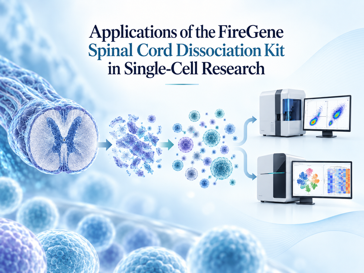
The FireGene Spinal Cord Dissociation Kit is designed to support the preparation of spinal cord tissue for downstream single-cell applications. By helping researchers generate a relatively uniform cell suspension while reducing aggregates and tissue fragments, the kit can support a range of experimental workflows, including single-cell RNA sequencing, flow cytometry, fluorescence-activated cell sorting, primary cell research, and studies of neuroinflammation and spinal cord injury.
1. Single-Cell RNA Sequencing of Spinal Cord Tissue
Single-cell RNA sequencing, or scRNA-seq, has become one of the most important technologies for studying cellular heterogeneity in nervous-system tissues.
Traditional bulk RNA sequencing measures gene expression across an entire sample. Although useful, bulk analysis can mask important differences between individual cell populations. In contrast, scRNA-seq enables researchers to examine gene-expression profiles at the level of individual cells.
In spinal cord research, single-cell sequencing can be used to characterize populations such as:
excitatory and inhibitory neurons;
astrocytes;
oligodendrocytes;
oligodendrocyte precursor cells;
microglia;
endothelial cells;
perivascular cells;
infiltrating immune cells;
and other specialized cellular subsets.
The quality of the cell suspension is especially important for single-cell sequencing. Excessive cellular aggregation can interfere with droplet-based partitioning, while large amounts of debris may reduce effective cell recovery. Damaged cells can also release RNA into the extracellular environment, potentially increasing background signals.
The FireGene Spinal Cord Dissociation Kit can therefore serve as an upstream sample-preparation tool for researchers developing spinal cord scRNA-seq workflows.
After dissociation, researchers may evaluate cell concentration, viability, aggregation, and debris levels before proceeding to library preparation. Depending on the experimental objective, additional enrichment, filtration, or dead-cell removal steps may also be incorporated.
2. Flow Cytometry and Cellular Phenotyping
Flow cytometry is widely used to analyze the cellular composition of spinal cord samples.
Once spinal cord tissue has been converted into a single-cell suspension, cells can be labeled with fluorescent antibodies or other markers and analyzed according to parameters such as surface protein expression, intracellular markers, viability, and cell size.
Flow cytometric analysis is particularly useful in studies involving glial and immune cell populations.
For example, researchers may investigate:
microglial populations;
infiltrating monocytes;
macrophages;
lymphocyte populations;
endothelial cells;
astrocyte-associated markers;
and other defined cellular subsets.
Spinal cord inflammation is often accompanied by substantial changes in immune-cell composition. Flow cytometry allows researchers to quantify these changes and compare them across experimental groups.
Reliable dissociation is important because flow cytometers are designed to analyze individual suspended particles. Cellular clumps may result in inaccurate event detection or instrument blockage, while incomplete tissue digestion can reduce the recovery of specific cell populations.
A well-prepared single-cell suspension therefore improves the compatibility of spinal cord samples with cytometric analysis.
3. Fluorescence-Activated Cell Sorting
Fluorescence-activated cell sorting, or FACS, extends flow cytometry by allowing selected cells to be physically isolated from a heterogeneous population.
This approach is particularly valuable when researchers need purified spinal cord cell populations for downstream molecular or functional studies.
For example, specific populations may be isolated for:
RNA extraction;
gene-expression analysis;
protein analysis;
cell culture;
sequencing;
functional assays;
or comparative molecular profiling.
Researchers studying neuroimmune interactions may isolate microglia or infiltrating immune cells, while studies focused on neural support cells may enrich for astrocytes or oligodendrocyte-lineage populations.
The success of FACS depends strongly on sample quality. Aggregates can interfere with sorting accuracy, while cellular debris may complicate gating strategies. Excessively damaged cells can also reduce recovery of viable target populations.
For this reason, tissue dissociation represents an important upstream component of a successful spinal cord FACS workflow.
4. Primary Cell Isolation and Culture
Primary spinal cord-derived cells are frequently used in experimental neuroscience.
Unlike immortalized cell lines, primary cells retain many biological characteristics associated with their tissue of origin. They can therefore provide useful experimental models for investigating cellular signaling, cell–cell interactions, stress responses, and molecular pathways.
Following tissue dissociation, researchers may obtain mixed cellular populations or further enrich selected cell types.
Potential experimental applications include research involving:
primary glial cells;
microglial populations;
neural cell populations;
mixed spinal cord cultures;
and other tissue-derived cellular preparations.
Primary cell experiments may investigate responses to cytokines, growth factors, signaling molecules, experimental compounds, or changes in culture conditions.
However, successful primary cell preparation requires careful control of the dissociation process. Excessive enzymatic digestion or mechanical force may reduce cell viability, while insufficient dissociation may leave tissue fragments and aggregates.
The FireGene Spinal Cord Dissociation Kit provides researchers with a standardized starting point for preparing spinal cord tissue before subsequent culture or enrichment procedures.
5. Neuroinflammation Research
Neuroinflammation is a major area of spinal cord research.
Microglia and other immune-associated cell populations respond to tissue injury, infection, cellular stress, and changes in the local microenvironment. Peripheral immune cells may also enter spinal cord tissue under certain experimental conditions.
Single-cell preparation enables researchers to investigate these populations individually.
For example, dissociated spinal cord cells may be analyzed to examine:
changes in microglial abundance;
inflammatory cell infiltration;
immune-cell phenotypes;
cytokine-associated signaling;
activation-related markers;
transcriptional changes;
and interactions between neural and immune populations.
Single-cell sequencing has further demonstrated that microglia and infiltrating immune cells may contain multiple molecularly distinct subpopulations.
Because these differences can be obscured in bulk tissue analysis, single-cell approaches are particularly valuable for studying neuroinflammatory biology.
Efficient spinal cord dissociation therefore provides an important technical foundation for studying the cellular complexity of inflammatory responses.
6. Spinal Cord Injury Research
Spinal cord injury can produce substantial changes in tissue architecture, cellular composition, inflammatory signaling, and neural function.
Following experimental injury, the spinal cord may contain damaged neural cells, activated glia, infiltrating immune cells, vascular-associated cells, and dynamically changing cellular populations.
Single-cell technologies allow researchers to examine how these populations respond over time.
Potential applications include investigation of:
acute cellular responses after injury;
inflammatory-cell recruitment;
microglial activation;
astrocyte responses;
oligodendrocyte-lineage changes;
vascular remodeling;
cellular stress pathways;
and tissue repair-associated processes.
Researchers may collect spinal cord samples at different time points after injury and compare their cellular composition using scRNA-seq or flow cytometry.
This type of experimental design can help identify time-dependent transitions that would be difficult to detect using bulk tissue analysis alone.
Consistent dissociation protocols are especially important in such studies because injured tissue may differ substantially from untreated spinal cord tissue in terms of structural integrity, extracellular material, and cellular composition.
7. Neurodegeneration and Neurological Disease Models
Single-cell analysis is also increasingly applied to experimental models of neurological and neurodegenerative disorders involving the spinal cord.
Researchers may investigate changes in neuronal populations, glial-cell states, immune responses, and gene-expression programs.
Potential research areas include:
motor neuron biology;
neurodegenerative disease models;
demyelination and remyelination;
neuroimmune signaling;
axonal injury;
cellular stress;
and glial dysfunction.
By comparing control and experimental samples, researchers can identify cell-type-specific molecular alterations.
For example, a transcriptional pathway that appears only moderately altered in whole-tissue RNA analysis may show a pronounced change within a specific microglial or neuronal subpopulation when examined using single-cell methods.
This improved cellular resolution is one of the principal advantages of modern single-cell neuroscience.
8. Cell Population Profiling and Comparative Studies
Another important application of spinal cord dissociation is cellular population profiling.
Researchers often need to determine whether experimental conditions alter the relative abundance or phenotype of specific cell types.
A general workflow may involve:
Spinal Cord Collection → Tissue Dissociation → Filtration → Cell Quality Assessment → Flow Cytometry or Single-Cell Sequencing → Cell Population Analysis
Such workflows can be used to compare:
control and experimental groups;
different developmental stages;
different anatomical regions;
treated and untreated samples;
different genetic backgrounds;
or multiple experimental time points.
Standardized dissociation can help reduce sample-preparation variability, which is particularly important when quantitative comparisons are made between experimental groups.
Conclusion
The preparation of a reliable single-cell suspension is a fundamental step in many modern spinal cord research workflows.
The FireGene Spinal Cord Dissociation Kit can support researchers working with spinal cord tissue in applications including single-cell RNA sequencing, flow cytometry, fluorescence-activated cell sorting, primary cell research, neuroinflammation studies, spinal cord injury models, neurological disease research, and cell population profiling.
As neuroscience increasingly moves toward cell-type-specific and single-cell approaches, sample preparation becomes increasingly important. The quality of tissue dissociation can influence cell recovery, viability, aggregation, debris levels, and the reliability of downstream analysis.
By providing a practical approach for spinal cord single-cell preparation, the FireGene Spinal Cord Dissociation Kit can serve as an upstream component of experimental workflows designed to investigate the cellular and molecular complexity of spinal cord biology.


