
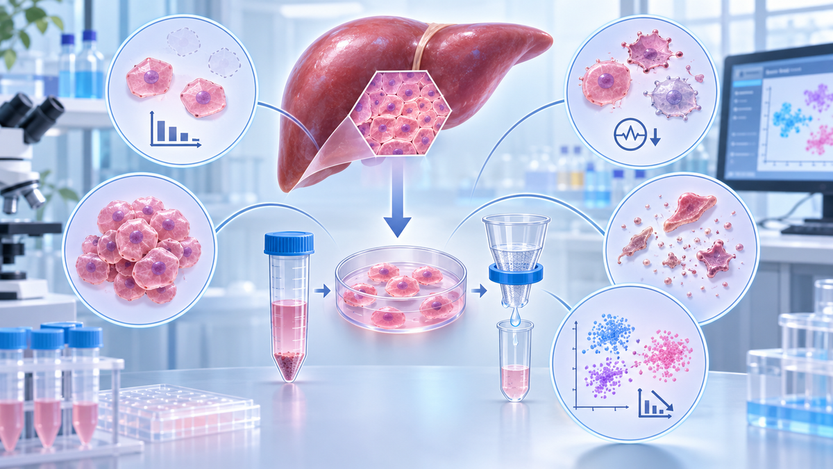
Liver cell isolation is an essential step in hepatic biology, liver disease research, drug metabolism studies, and single-cell RNA sequencing (scRNA-seq). However, obtaining viable, representative single-cell suspensions from liver tissue remains technically challenging. Common difficulties include low cell yield, reduced hepatocyte viability, cell aggregation, excessive debris, incomplete enzymatic digestion, and selective loss of specific hepatic cell populations. These problems can compromise downstream experiments, introduce cellular bias, and reduce data reproducibility. This article examines the most frequent problems encountered during liver cell isolation, explains their underlying causes, and presents practical troubleshooting approaches for improving tissue dissociation, cell recovery, and sample quality.
1. Introduction: Why Is Liver Cell Isolation Challenging?
The liver contains diverse cellular populations with distinct biological functions, structural characteristics, and sensitivities to experimental processing.
Hepatocytes account for a substantial proportion of liver tissue volume and perform essential metabolic functions. Non-parenchymal populations, including Kupffer cells, hepatic stellate cells, liver sinusoidal endothelial cells (LSECs), and immune cells, contribute to inflammation, extracellular matrix remodeling, and tissue homeostasis.
Successful liver cell isolation requires the disruption of extracellular matrix components and cell–cell interactions while preserving the integrity of individual cells.
However, the process can be affected by numerous variables, including tissue condition, enzymatic activity, digestion duration, mechanical stress, filtration, and centrifugation.
These challenges become particularly important in single-cell applications, where poor sample quality can introduce technical artifacts and alter the apparent representation of hepatic cell populations.
Understanding the causes of common isolation problems is therefore essential for optimizing experimental reproducibility.
2. Problem 1: Low Liver Cell Yield
2.1 Possible Causes
Low cell yield is among the most frequently encountered challenges during liver tissue dissociation.
One major cause is incomplete enzymatic digestion. Liver tissue contains extracellular matrix components, including collagen, laminin, and fibronectin, which maintain structural integrity.
If enzymatic activity is insufficient, cells may remain trapped within tissue fragments.
Additional causes include:
Inappropriate tissue fragment size
Insufficient or inconsistent digestion
Reduced enzyme activity during storage or preparation
Excessive cell loss during filtration and washing
Inappropriate centrifugation conditions
Poor initial tissue quality
Fibrotic and cirrhotic liver samples may be particularly difficult to dissociate because increased collagen deposition creates additional structural barriers.
2.2 Troubleshooting Strategies
Optimize enzymatic digestion. Evaluate digestion duration, enzyme activity, and tissue response according to the validated protocol.
Prepare uniform tissue fragments. Relatively consistent fragment sizes improve enzyme accessibility and reduce variability between samples.
Minimize processing losses. Avoid unnecessary filtration, repeated transfers, and excessive washing.
Evaluate tissue condition. Record sample mass, ischemic interval, storage conditions, and visible pathological changes.
Importantly, extending digestion is not always beneficial. Longer exposure may improve tissue breakdown but simultaneously reduce cell viability.
The objective should be to maximize viable cell recovery rather than total cell count alone.
3. Problem 2: Low Hepatocyte Viability
3.1 Why Hepatocytes Are Vulnerable
Hepatocytes are relatively large, metabolically active cells that are sensitive to mechanical injury and environmental changes.
During isolation, excessive shear stress, prolonged enzymatic treatment, or inappropriate centrifugation can disrupt cellular membranes.
Delayed tissue processing may also contribute to cellular damage through ischemic stress and metabolic deterioration.
Common indicators of reduced hepatocyte viability include membrane blebbing, irregular morphology, increased dead-cell staining, and excessive cellular debris.
3.2 Troubleshooting Strategies
Reduce mechanical stress. Use gentle resuspension techniques and consider wide-bore pipette tips when appropriate.
Avoid overdigestion. Monitor tissue dissociation and terminate digestion according to the validated procedure.
Maintain suitable sample conditions. Minimize delays and follow the recommended temperature and buffer requirements for each processing stage.
Optimize centrifugation. Select conditions appropriate for the target cell population rather than applying a universal protocol to all hepatic cells.
Assess viability promptly. Evaluate cell viability immediately after isolation and before downstream applications.
Trypan blue exclusion and fluorescence-based viability assays are commonly used for initial assessment.
However, viability measurements should be interpreted alongside cell recovery and functional indicators.
For primary hepatocyte culture, attachment efficiency, cellular morphology, and metabolic activity may provide additional information about cell quality.
4. Problem 3: Cell Clumping and Aggregation
4.1 Why Do Liver Cells Form Aggregates?
Cell aggregation can substantially compromise liver cell preparation.
One important cause is the release of extracellular DNA from damaged or lysed cells.
Extracellular DNA can increase suspension viscosity and promote interactions between cells and tissue debris.
Incomplete enzymatic digestion may also leave residual cell clusters and extracellular matrix fragments.
Additional contributing factors include prolonged sample handling, inappropriate cell concentration, and insufficient suspension mixing.
For droplet-based scRNA-seq, cellular aggregates are particularly problematic because they can interfere with microfluidic processing and increase multiplet formation.
4.2 Troubleshooting Strategies
Improve digestion consistency. Ensure tissue is sufficiently dissociated before proceeding to filtration.
Minimize cell damage. Gentle processing reduces the release of intracellular contents.
Consider DNase treatment. When compatible with the experimental workflow, DNase may help reduce aggregation associated with extracellular DNA.
Use appropriate filtration. Select cell strainers based on target cell size and downstream requirements.
Avoid prolonged storage of suspensions. Process isolated cells promptly whenever practical.
Researchers should exercise caution when selecting filtration pore size.
Mature hepatocytes may be larger than many non-parenchymal cells, and excessively restrictive filtration can reduce hepatocyte recovery.
Therefore, cell aggregation control should not come at the expense of unnecessary cell-type depletion.
5. Problem 4: Excessive Cellular Debris and Dead Cells
5.1 Possible Causes
Excessive debris often indicates cellular damage or incomplete tissue processing.
Common sources include:
Overdigestion of liver tissue
Forceful pipetting or mechanical disruption
Poor sample preservation
Necrotic or damaged tissue regions
Inappropriate centrifugation and resuspension
Dead cells may release intracellular RNA and other molecular components into the suspension.
In scRNA-seq experiments, extracellular RNA can contribute to ambient RNA contamination, complicating cell-type identification and gene expression analysis.
5.2 Troubleshooting Strategies
Begin by evaluating the condition of the original tissue and identifying the processing stage associated with increased cell damage.
Gentle mechanical handling and appropriate enzymatic treatment can help minimize debris generation.
If excessive debris remains, researchers may consider additional cleanup methods, including appropriate filtration, density-based separation, or dead-cell removal procedures.
However, cleanup steps may introduce cell loss and alter the representation of different hepatic cell populations.
Therefore, the benefits of debris removal must be balanced against potential losses of viable cells.
For low-input samples, reducing unnecessary handling may be more beneficial than performing multiple purification steps.
6. Problem 5: Incomplete Liver Tissue Digestion
Incomplete digestion may be identified by persistent tissue fragments, visible cell clusters, or difficulty generating a homogeneous suspension.
The problem is particularly relevant to fibrotic liver tissue, where increased extracellular matrix deposition can restrict enzymatic access.
Possible Causes
Inadequate enzyme activity, inconsistent fragment preparation, insufficient mixing, and tissue-specific matrix composition can all contribute to poor digestion.
Different tissue sources may also require different handling strategies.
For example, healthy liver tissue and heavily fibrotic liver tissue may not respond identically to the same digestion conditions.
Recommended Solutions
Researchers should verify enzyme preparation and storage, assess tissue fragment size, and monitor the progress of dissociation.
When additional digestion is required, its effect on cell viability should be evaluated.
Aggressive mechanical disruption should not be used as a substitute for inadequate enzymatic digestion because it may cause extensive cell damage.
7. Problem 6: Uneven Recovery of Hepatic Cell Populations
A major challenge in hepatic cell profiling is achieving representative recovery of different cell populations.
Hepatocytes, Kupffer cells, hepatic stellate cells, endothelial cells, and cholangiocytes differ in size, density, structural interactions, and sensitivity to tissue processing.
Consequently, isolation conditions that favor one population may reduce the recovery of another.
For example, centrifugation conditions designed to enrich hepatocytes may selectively reduce the proportion of smaller non-parenchymal cells in the recovered fraction.
Likewise, mechanical stress may preferentially damage fragile cell populations.
Troubleshooting Strategies
Researchers should select isolation conditions based on their experimental objectives.
For whole-liver scRNA-seq, maintaining cellular diversity is often a central priority.
For primary hepatocyte culture, viable hepatocyte recovery and functional integrity may be more important.
Cell-type representation can be evaluated using flow cytometry, immunostaining, or transcriptomic markers.
Useful examples include:
|
Cell Population |
Representative Research Markers |
|
Hepatocytes |
ALB, HNF4A, CYP enzymes |
|
Kupffer cells |
CD68, CLEC4F, TIMD4 |
|
Hepatic stellate cells |
LRAT, DES, RGS5 |
|
Liver sinusoidal endothelial cells |
STAB2, LYVE1, CLEC4G |
|
Cholangiocytes |
KRT7, KRT19, EPCAM |
Marker expression depends on species, cell state, and disease context. Multiple markers are generally preferable to a single marker for reliable characterization.
8. Problem 7: Poor Single-Cell RNA Sequencing Data Quality
Even when liver cell isolation produces an apparently adequate cell suspension, downstream sequencing quality may remain suboptimal.
Potential problems include high ambient RNA levels, increased mitochondrial RNA fractions, cellular multiplets, and stress-associated gene expression.
Tissue dissociation can also induce transcriptional changes that complicate interpretation of biological differences between experimental groups.
Troubleshooting Strategies
Researchers should evaluate cell viability, aggregate frequency, and suspension cleanliness before sequencing.
Consistent tissue processing conditions are particularly important when comparing healthy and diseased liver samples.
Additional strategies include minimizing processing delays, reducing unnecessary mechanical manipulation, and standardizing digestion conditions.
When intact cell recovery is persistently problematic, single-nucleus RNA sequencing (snRNA-seq) may provide an alternative for selected research questions.
However, scRNA-seq and snRNA-seq capture different transcriptomic compartments and may exhibit distinct cell-type representation biases.
The choice of method should therefore reflect the biological objectives of the study.
9. Quick Troubleshooting Guide for Liver Cell Isolation
|
Problem |
Likely Cause |
Recommended Solution |
|
Low cell yield |
Incomplete digestion |
Optimize enzymatic digestion and tissue preparation |
|
Low hepatocyte viability |
Mechanical stress or overdigestion |
Reduce handling intensity and reassess digestion duration |
|
Cell aggregation |
Extracellular DNA and residual tissue fragments |
Minimize lysis and evaluate compatible DNase treatment |
|
Excessive debris |
Cell damage or poor tissue quality |
Improve handling and consider selective cleanup |
|
Incomplete digestion |
Dense extracellular matrix or low enzyme activity |
Verify enzyme performance and tissue-specific conditions |
|
Uneven cell recovery |
Cell-type-specific losses |
Optimize filtration and separation strategy |
|
Poor scRNA-seq quality |
Dead cells, aggregates, or ambient RNA |
Strengthen quality control before library preparation |
10. Supporting Reliable Liver Cell Preparation with FireGene
Reliable liver cell isolation begins with appropriate tissue dissociation reagents and a consistent experimental workflow.
The FireGene Liver Dissociation Kit for Hepatic Cell Profiling is designed to support the preparation of single-cell suspensions from liver tissue for downstream cellular research.
Its liver-oriented dissociation solution provides researchers with a reagent system for hepatic tissue processing.
Potential applications include single-cell RNA sequencing, hepatic cell profiling, cell culture, and other cell-based analyses.
By incorporating a standardized dissociation reagent into an optimized workflow, researchers can seek to reduce reagent-preparation variability and improve experimental consistency.
However, final cell yield, viability, and cell-type representation depend on tissue condition, handling procedures, and downstream processing. Performance should be validated for the specific experimental model.
Explore the FireGene Liver Dissociation Kit:
https://firegene.com/products/liver-dissociation-kit-fg-ba3323
11. Frequently Asked Questions
Why is my liver cell yield low after dissociation?
Low cell yield may result from incomplete digestion, insufficient enzyme activity, poor tissue quality, or cell loss during filtration and centrifugation. Reviewing each stage of the isolation workflow can help identify the main cause.
How can I improve hepatocyte viability during isolation?
Minimize tissue processing delays, avoid excessive mechanical force, optimize digestion conditions, and use suitable washing and centrifugation procedures.
What causes liver cells to clump after dissociation?
Cell aggregation is frequently associated with extracellular DNA released from damaged cells, residual matrix components, and incomplete tissue dissociation.
Should dead cells always be removed before scRNA-seq?
Dead-cell removal can improve suspension cleanliness, but it may also reduce total cell recovery. The decision should depend on sample viability, available cell numbers, and the sequencing platform's requirements.
Can the same dissociation method be used for healthy and fibrotic liver tissue?
The same general workflow may serve as a starting point, but fibrotic tissue often requires additional optimization because of differences in extracellular matrix composition and tissue structure.
12. Conclusion
Liver cell isolation involves multiple interconnected challenges, including low cell yield, reduced hepatocyte viability, aggregation, debris formation, and unequal recovery of hepatic cell populations.
Addressing these problems requires more than simply increasing digestion intensity.
A successful workflow combines appropriate enzymatic treatment, gentle mechanical handling, controlled sample processing, and application-specific quality assessment.
The FireGene Liver Dissociation Kit for Hepatic Cell Profiling provides a liver-focused reagent option that can be incorporated into such workflows.
Ultimately, optimizing the complete tissue-to-cell preparation process is essential for generating reliable samples for hepatic cell profiling, flow cytometry, primary cell research, and single-cell transcriptomics.
For Research Use Only. Not for diagnostic or therapeutic use.


