
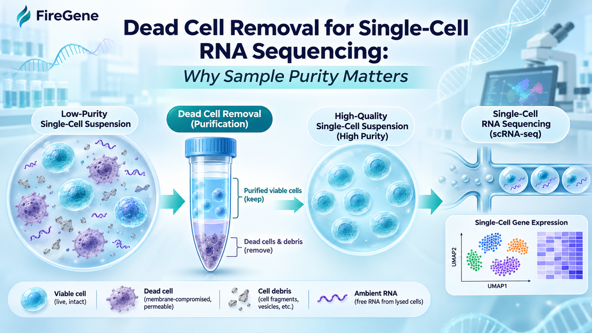
Single-cell RNA sequencing (scRNA-seq) has become an essential technology for investigating cellular heterogeneity, gene-expression patterns, and complex biological systems. However, reliable single-cell transcriptomic analysis depends heavily on the quality of the starting cell suspension. Dead cells, cellular debris, and extracellular RNA can interfere with cell capture, increase background contamination, and compromise downstream data interpretation. During tissue dissociation, cryopreservation, and sample handling, damaged cells may release intracellular RNA into the surrounding environment, contributing to ambient RNA contamination. Effective dead cell removal can enrich viable-cell populations, improve suspension uniformity, and reduce certain sources of experimental variability. Nevertheless, purification procedures must be carefully optimized to minimize viable-cell loss and avoid changes in cellular composition. This article examines the effects of dead cell contamination on scRNA-seq, reviews commonly used removal methods, and discusses strategies for improving sample purity, cell recovery, and sequencing reliability.
1. Introduction
Single-cell RNA sequencing enables researchers to characterize gene-expression profiles at the resolution of individual cells. Unlike bulk RNA sequencing, which measures average transcript abundance across a population, scRNA-seq can reveal cellular heterogeneity, identify rare populations, and characterize distinct transcriptional states.
This capability has made scRNA-seq particularly valuable in cancer research, immunology, developmental biology, neuroscience, and tissue microenvironment studies.
Despite these advantages, the reliability of single-cell sequencing depends strongly on the quality of the biological material entering the workflow.
A suitable single-cell suspension should contain predominantly viable, intact, individual cells with minimal aggregates and unwanted cellular material.
However, samples prepared from solid tissues, cryopreserved specimens, and fragile primary cells often contain substantial amounts of dead cells and debris.
These contaminants may interfere with cell counting, microfluidic loading, transcript capture, and downstream bioinformatic analysis.
Furthermore, dead cells can release intracellular RNA into the surrounding suspension, introducing extracellular transcripts that may be captured during sequencing.
Consequently, dead cell removal is an important consideration in scRNA-seq sample preparation, particularly when the starting material exhibits reduced viability or extensive cellular damage.
2. Sources of Dead Cells and Cellular Debris in scRNA-seq Samples
Dead cells and cellular debris may originate from multiple stages of sample preparation.
2.1 Tissue Dissociation
Solid tissues must generally be converted into single-cell suspensions before conventional scRNA-seq processing.
This commonly requires enzymatic digestion, mechanical disruption, or a combination of both approaches.
Although these procedures facilitate cell isolation, excessive digestion or aggressive mechanical handling may damage cell membranes and compromise viability.
Prolonged exposure to dissociation enzymes can also induce stress responses and alter gene-expression profiles.
The susceptibility of cells to dissociation-related damage varies among tissues and cellular populations.
2.2 Cryopreservation and Thawing
Cryopreserved specimens provide flexibility for sample transportation, storage, and experimental scheduling.
However, freezing and thawing can introduce osmotic stress, membrane damage, and cellular injury.
Certain primary cell populations are particularly sensitive to cryopreservation-associated damage.
Following thawing, the suspension may contain intact viable cells, nonviable cells, membrane fragments, and soluble intracellular components.
The proportion of these contaminants depends on the preservation procedure, storage conditions, and thawing method.
2.3 Sample Transportation and Handling
Delays between tissue collection and processing may contribute to cellular stress and reduced viability.
Temperature fluctuations, prolonged storage, repeated centrifugation, and excessive pipetting may further compromise cell integrity.
Appropriate handling conditions are therefore important for preserving sample quality before sequencing.
2.4 Biological Characteristics of the Starting Material
Some biological samples naturally contain substantial amounts of nonviable cellular material.
Necrotic tumors, inflamed tissues, and damaged organs may contain dead cells even before laboratory processing begins.
These samples can present additional challenges because extracellular matrix fragments, cellular aggregates, and heterogeneous cell populations may complicate purification.
3. How Dead Cells Compromise scRNA-seq Data Quality
3.1 Ambient RNA Contamination
Ambient RNA contamination is one of the major concerns associated with dead cells in single-cell sequencing.
When cell membranes lose integrity, intracellular RNA can escape into the surrounding medium.
During droplet-based scRNA-seq, this extracellular RNA may enter droplets containing viable cells or droplets without intact cells.
As a result, sequencing libraries may contain transcripts that do not originate from the captured cell.
Ambient RNA contamination can introduce several analytical problems, including:
Increased background transcript detection
False identification of cell-type-specific markers
Reduced specificity of gene-expression profiles
Difficulty distinguishing related cellular populations
Misinterpretation of biological pathways and cellular states
For example, RNA released from damaged epithelial cells may be detected in droplets containing immune cells, potentially creating misleading expression patterns.
Young and Behjati (2020) described the effects of ambient RNA contamination and introduced SoupX as a computational approach for estimating and reducing background contamination in droplet-based scRNA-seq datasets.
Although computational correction can reduce certain contamination effects, preventing excessive RNA release during sample preparation remains important.
3.2 Reduced Cell Capture Efficiency
Dead cell contamination may interfere with accurate sample counting and loading.
Single-cell sequencing platforms generally require cell suspensions within specified concentration ranges.
If a substantial proportion of counted cells is nonviable, the actual number of intact viable cells available for capture may be lower than expected.
This can reduce the number of high-quality cells recovered after sequencing.
Aggregates and cellular fragments may also complicate loading and contribute to technical variability.
Therefore, accurate viable-cell counting and appropriate suspension cleanup are important for consistent sample preparation.
3.3 Low-Quality Transcriptomic Profiles
Damaged or dying cells may produce transcriptomic profiles that differ substantially from those of healthy cells.
Such profiles can exhibit reduced transcript complexity, altered expression of stress-associated genes, or elevated mitochondrial RNA proportions.
However, these indicators are not universally specific for dead cells.
For example, mitochondrial transcript abundance can vary naturally among tissues, cell types, and metabolic states.
Quality-control thresholds should therefore be selected according to the experimental system rather than applying identical filtering criteria to every dataset.
3.4 Increased Cell Aggregation
Cell damage can release genomic DNA and other intracellular components that promote cell aggregation.
Cell clumps may interfere with microfluidic processing and increase the likelihood of capturing more than one cell within a partition.
These multiplets can complicate downstream analysis by generating mixed transcriptomic profiles.
Removing damaged cells, reducing extracellular DNA, and maintaining appropriate cell concentrations can help improve suspension uniformity.
4. Cell Viability and Sample Purity Requirements
Cell viability is a widely used indicator of sample quality in scRNA-seq preparation.
It represents the proportion of viable cells among the total counted cell population.
Cell Viability (%) = (Viable Cells / Total Cells) × 100
For example, a suspension containing 850,000 viable cells and 150,000 nonviable cells has a viability of 85%.
High viability is generally desirable for conventional droplet-based single-cell sequencing.
Some 10x Genomics sample preparation guidance recommends targeting viability above 90%, although acceptable viability depends on the platform, assay chemistry, and sample characteristics.
A practical interpretation of viability measurements is summarized below.
|
Cell Viability |
Sample Condition |
General Consideration |
|
≥90% |
High viability |
Additional purification may be unnecessary |
|
80–90% |
Relatively high viability |
Evaluate debris and aggregation |
|
70–80% |
Moderate viability |
Consider whether dead cell removal would be beneficial |
|
Below 70% |
Low viability |
Assess purification options and sample suitability |
These ranges are illustrative rather than universal acceptance criteria.
Importantly, cell viability alone does not fully describe sample quality.
A suspension may contain a high percentage of intact cells while still exhibiting substantial extracellular RNA contamination or cellular aggregation.
Therefore, researchers should assess viability together with cell concentration, debris abundance, aggregation, and viable-cell recovery.
5. Common Dead Cell Removal Methods for scRNA-seq
Several purification methods can improve suspension quality before sequencing.
5.1 Filtration
Filtration is commonly performed after tissue dissociation to remove undissociated fragments and large aggregates.
Cell strainers with pore sizes such as 40 µm or 70 µm are frequently used, depending on the target cell population.
This approach improves suspension uniformity and may reduce microfluidic obstruction.
However, filtration cannot reliably distinguish viable cells from nonviable cells of similar size.
Consequently, additional purification may be necessary when substantial dead cell contamination is present.
5.2 Differential Centrifugation
Differential centrifugation separates suspended material according to sedimentation characteristics.
Under suitable conditions, intact cells can be pelleted while some smaller debris remains in the supernatant.
Although this approach is straightforward and widely accessible, it often provides limited discrimination between viable and nonviable cells.
Repeated washing may also increase viable-cell loss.
5.3 Density Gradient Centrifugation
Density gradient centrifugation separates cellular components according to their buoyant density and other physical characteristics.
During centrifugation, different populations distribute within the separation medium, allowing researchers to recover a fraction enriched in viable cells.
This approach can be useful for tissue-derived suspensions containing mixtures of intact cells, damaged cells, and cellular debris.
The effectiveness of density-based purification depends on the separation medium, centrifugation conditions, starting sample composition, and fraction collection procedure.
Because different cell types may exhibit overlapping densities, complete removal of nonviable cells cannot be assumed.
5.4 Magnetic-Based Dead Cell Removal
Magnetic separation uses reagents that selectively recognize unwanted cellular populations.
Depending on the system, dead or apoptotic cells may be labeled and retained during magnetic separation.
The remaining fraction can then be collected for downstream analysis.
Magnetic depletion may provide effective viable-cell enrichment but requires additional reagents and processing steps.
Researchers should also evaluate whether the method preferentially removes fragile or biologically important cell populations.
5.5 Fluorescence-Activated Cell Sorting
FACS enables the selection of viable cells based on fluorescent markers and light-scattering characteristics.
Viability dyes can identify many cells with compromised membranes, allowing these populations to be excluded.
FACS can provide highly selective purification, particularly when specific cell populations are required.
However, sorting can introduce additional processing time, mechanical stress, and potential cell loss.
6. Strategies for Improving Sample Purity Before Sequencing
6.1 Optimize Tissue Dissociation Conditions
Reducing cellular damage during dissociation is one of the most effective ways to limit subsequent contamination.
Enzyme concentration, digestion duration, temperature, and mechanical handling should be adapted to the tissue being processed.
Gentle procedures can help preserve fragile populations and reduce the generation of cellular debris.
6.2 Minimize Processing Time
Extended handling may increase cell stress and death.
Samples should be processed efficiently under conditions appropriate for the target cell type and sequencing workflow.
Unnecessary waiting periods and repeated centrifugation should be avoided whenever possible.
6.3 Control Cell Concentration
Excessively concentrated suspensions may promote aggregation and complicate accurate cell counting.
Conversely, highly diluted samples can increase handling-related losses.
Maintaining an appropriate concentration supports more consistent purification and subsequent sequencing preparation.
6.4 Reduce Cell Clumping
Extracellular DNA released from damaged cells can contribute to aggregation.
Where compatible with the sample, DNase treatment may reduce DNA-mediated clumping.
Gentle mixing and appropriate buffer composition can further improve suspension uniformity.
6.5 Balance Viability and Cell Recovery
Purification should not focus exclusively on maximizing the percentage of viable cells.
Every separation step introduces some risk of losing viable cells.
Viable-cell recovery can be calculated as:
Viable-Cell Recovery (%) = (Viable Cells After Purification / Viable Cells Before Purification) × 100
A purification method that produces high final viability but removes most of the viable starting cells may be unsuitable for limited tissue specimens or rare-cell studies.
The optimal strategy should preserve sufficient cell numbers while improving sample quality.
7. Can Dead Cell Removal Improve scRNA-seq Results?
Dead cell removal can improve certain aspects of scRNA-seq sample preparation, particularly when the starting suspension contains substantial nonviable material.
Removing damaged cells before additional lysis may reduce sources of further extracellular RNA release.
Reducing aggregates and particulate contamination may also improve sample loading consistency.
Furthermore, enrichment of viable cells can improve the accuracy of cell concentration measurements and the proportion of intact cells entering the sequencing workflow.
Nevertheless, purification cannot guarantee complete removal of ambient RNA.
Extracellular transcripts released before cleanup may remain in the suspension, even when visible debris has been reduced.
Computational approaches such as SoupX and CellBender can complement experimental purification by identifying or correcting certain forms of ambient RNA contamination.
However, computational correction cannot recover lost cells or fully reverse biological changes introduced during sample processing.
Effective quality control should therefore combine appropriate sample preparation, experimental assessment, and downstream bioinformatic analysis.
8. Limitations of Dead Cell Removal
Although purification can improve sample quality, it may also introduce experimental bias.
Different cell populations vary in size, density, membrane integrity, and sensitivity to handling.
Certain fragile or stressed populations may be disproportionately lost during purification.
This can alter the representation of cell types in the final sample.
In tumor microenvironment studies, for example, excessive purification may affect the recovery of fragile stromal cells or stressed tumor-associated populations.
Similarly, filtration can exclude larger cells, while density-based separation may favor populations with particular physical properties.
Denisenko et al. (2020) demonstrated that tissue dissociation and storage procedures can influence both recovered cell composition and gene-expression profiles.
Therefore, researchers should evaluate whether purification introduces systematic changes that could affect biological interpretation.
In experiments involving rare populations or highly heterogeneous specimens, comparing purified and unpurified samples may help identify preparation-related bias.
9. FireGene Dead Cell Debris & Impurities Removal Kit for scRNA-seq Sample Preparation
The FireGene Dead Cell Debris & Impurities Removal Kit (Cat. No. FG-BA3339) is designed to support the purification of single-cell suspensions by reducing dead cells, cellular debris, and other impurities.
The kit employs a density-based separation approach intended to enrich viable cells from mixed cellular suspensions.
This strategy may be useful when preparing samples generated through tissue dissociation, cell harvesting, or other workflows in which cellular damage produces unwanted material.
For scRNA-seq applications, purification may help improve suspension cleanliness before cell counting and platform loading.
Potential benefits include reducing the nonviable fraction, decreasing particulate contamination, and improving the consistency of sample preparation.
However, the actual improvement in viability, debris removal, and cell recovery depends on the properties of the starting sample and experimental conditions.
Researchers should follow the manufacturer's recommended protocol and evaluate the recovered cells using appropriate quality-control measurements.
Product Name: FireGene Dead Cell Debris & Impurities Removal Kit
Catalog Number: FG-BA3339
Separation Principle: Density-based centrifugation
Application: Single-cell suspension purification for downstream research
Product Page: https://firegene.com/products/dead-cell-debris-removal-kit-fg-ba3339
The product is intended for research use only.
10. Conclusion
Dead cell contamination represents an important challenge in single-cell RNA sequencing.
Damaged cells can release intracellular RNA, generate cellular debris, contribute to aggregation, and reduce the quality of recovered single-cell transcriptomic profiles.
Removing dead cells before sequencing may improve the proportion of intact cells and reduce certain sources of experimental contamination.
Several methods are available, including filtration, differential centrifugation, density gradient separation, magnetic depletion, and fluorescence-activated cell sorting.
The appropriate purification strategy depends on the starting sample, required cell recovery, and downstream sequencing workflow.
Importantly, purification should be optimized to avoid unnecessary cell loss and changes in cellular composition.
For challenging samples containing substantial nonviable material, density-based approaches such as the FireGene Dead Cell Debris & Impurities Removal Kit provide an option for improving single-cell suspension quality.
By combining appropriate purification with viability assessment, cell recovery measurements, and downstream quality control, researchers can improve the consistency and interpretability of scRNA-seq experiments.


