
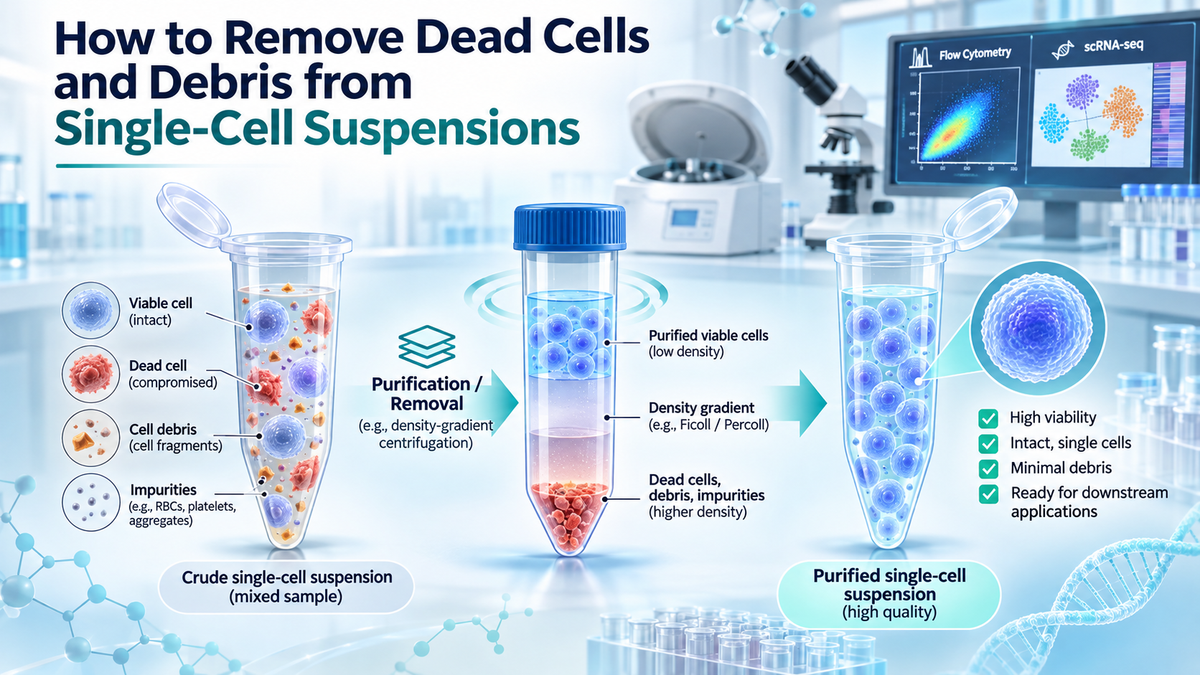
The preparation of high-quality single-cell suspensions is essential for reliable cellular and molecular research. However, tissue dissociation, cell harvesting, cryopreservation, and sample handling can generate dead cells, cellular fragments, and other impurities that compromise sample quality. These contaminants may interfere with single-cell RNA sequencing (scRNA-seq), flow cytometry, fluorescence-activated cell sorting (FACS), and primary cell culture. Effective removal strategies include filtration, differential centrifugation, density gradient separation, magnetic-based depletion, and fluorescence-based cell sorting. This article discusses the causes and consequences of cellular contamination, compares commonly used purification techniques, and outlines practical approaches for improving sample viability, purity, and recovery while preserving the biological characteristics of target cells.
1. Introduction: The Importance of Clean Single-Cell Suspensions
Single-cell technologies have transformed biomedical research by enabling scientists to investigate cellular heterogeneity, identify rare populations, and characterize biological processes at unprecedented resolution.
Applications such as single-cell RNA sequencing, flow cytometry, and primary cell culture depend heavily on the quality of the starting cell suspension.
Ideally, a single-cell suspension should contain a high proportion of viable, intact cells with minimal cellular debris, aggregates, and extracellular contaminants.
In practice, obtaining such suspensions can be challenging, particularly when processing solid tissues, frozen specimens, or samples containing substantial numbers of damaged cells.
During tissue dissociation, mechanical disruption and enzymatic digestion can compromise cell membranes. Similarly, cryopreservation, thawing, and prolonged sample handling may induce cellular stress and death.
These processes generate a mixture of intact viable cells, nonviable cells, apoptotic bodies, membrane fragments, and other impurities.
If these contaminants are not adequately managed, they can influence downstream experimental results.
For example, damaged cells may release intracellular RNA into the surrounding suspension, contributing to ambient RNA contamination during scRNA-seq. In flow cytometry, debris may produce unwanted events and complicate population identification.
Consequently, dead cell and debris removal is an important component of sample preparation, particularly when the initial suspension has low viability or contains substantial cellular contamination.
2. Sources and Effects of Dead Cells and Cellular Debris
2.1 Major Sources of Cellular Contamination
Dead cells and debris can accumulate during several stages of sample preparation.
Enzymatic tissue dissociation
Enzymes used to disrupt extracellular matrix components facilitate the release of individual cells from solid tissues. However, excessive digestion times, unsuitable enzyme concentrations, or inappropriate incubation conditions may compromise cell integrity.
Mechanical disruption
Tissue mincing, pipetting, homogenization, and filtration can expose cells to mechanical stress. Aggressive processing may damage cell membranes and generate cellular fragments.
Cryopreservation and thawing
Freezing and thawing introduce osmotic and thermal stresses that may cause membrane injury and cell death. Cryopreserved primary cells can be particularly sensitive to these effects.
Sample transportation and storage
Prolonged processing delays, temperature fluctuations, and unsuitable storage conditions may gradually reduce cell viability.
Biological characteristics of the starting tissue
Some samples, including necrotic tumors and inflamed tissues, naturally contain substantial amounts of dead or damaged cellular material.
2.2 Effects on Single-Cell RNA Sequencing
Dead cells represent a significant challenge in scRNA-seq sample preparation.
When cell membranes lose integrity, intracellular RNA may enter the extracellular environment. This material can subsequently be captured during single-cell partitioning and library preparation.
Ambient RNA contamination may increase background gene-expression signals, complicate cell-type annotation, and obscure differences between biologically distinct populations.
Dead cells and cellular fragments may also contribute to low-quality droplets or partitions, reducing the proportion of useful sequencing data.
Although computational tools can help identify or correct certain forms of contamination, preventing excessive RNA release during sample preparation remains important.
2.3 Effects on Flow Cytometry and Cell Sorting
Cellular debris can interfere with flow cytometric measurements by producing events with low forward and side scatter.
Dead cells may also exhibit increased nonspecific binding or altered fluorescence characteristics, potentially affecting population identification.
In FACS applications, aggregates and debris can contribute to sorting interruptions or reduced instrument performance.
Appropriate sample cleanup, combined with viability staining and gating strategies, helps improve analytical consistency.
3. Common Methods for Dead Cell and Debris Removal
Several separation techniques can improve the quality of single-cell suspensions. Each method has advantages and limitations.
3.1 Filtration
Filtration is commonly used to remove large tissue fragments, aggregates, and undissociated material.
Cell strainers with pore sizes of approximately 40–70 µm are frequently used, depending on the target cell type and experimental application.
This approach is simple, relatively inexpensive, and compatible with many tissue dissociation workflows.
However, filtration cannot reliably separate viable cells from similarly sized dead cells, and small debris may pass through the filter.
Therefore, filtration is generally most useful as an initial cleanup step.
3.2 Differential Centrifugation
Differential centrifugation separates suspended material according to sedimentation characteristics, which depend on particle size, density, and centrifugal force.
Under suitable conditions, intact cells sediment while some smaller debris remains in the supernatant.
Removing the supernatant can reduce certain contaminants.
Nevertheless, differential centrifugation often provides limited discrimination between viable and nonviable cells.
Repeated centrifugation may also increase cell loss or damage fragile populations.
3.3 Density Gradient Centrifugation
Density gradient centrifugation is a useful approach for separating cellular populations with different buoyant densities.
During centrifugation, cells and other particles distribute according to their physical properties and the characteristics of the separation medium.
This process can allow recovery of a fraction enriched in viable cells while reducing dead cells, fragments, and other impurities.
A typical density-based purification procedure includes preparing the single-cell suspension, introducing the appropriate separation medium, centrifuging under specified conditions, collecting the desired fraction, and washing the recovered cells.
The effectiveness of this approach depends on medium composition, sample characteristics, centrifugation parameters, and fraction collection accuracy.
Density-based separation does not guarantee complete removal of every nonviable cell, because cell populations may exhibit overlapping physical properties.
3.4 Magnetic-Based Dead Cell Removal
Magnetic separation systems can selectively deplete unwanted cells using reagents that recognize particular cellular characteristics.
Some dead cell removal systems target features associated with compromised or apoptotic cells.
Following labeling, magnetically retained cells are separated from the remaining suspension.
Magnetic depletion can be useful when physical separation alone is insufficient.
However, the method may require additional reagents, specialized equipment, and optimization to avoid unnecessary loss of target cells.
3.5 Fluorescence-Activated Cell Sorting
FACS enables direct separation of cells based on fluorescence and light-scattering characteristics.
Viability dyes such as propidium iodide and 7-AAD can identify many cells with compromised membranes, allowing these populations to be excluded during sorting.
FACS can provide highly selective purification, particularly when researchers need specific cell populations.
However, sorting can be time-consuming and may introduce additional handling stress.
For samples with extensive debris contamination, preliminary cleanup may improve sorting efficiency.
4. General Workflow for Removing Dead Cells and Debris
A well-designed purification workflow should improve suspension quality without unnecessarily compromising viable-cell recovery.
The following steps describe a general strategy. Experimental conditions should be adapted to the tissue, cell type, purification reagent, and downstream application.
Step 1: Prepare the Starting Suspension
Generate a single-cell suspension using a suitable tissue dissociation or cell harvesting method.
Maintain cells in an appropriate buffer or medium and minimize unnecessary processing delays.
Gentle handling is especially important for fragile primary cells.
Step 2: Remove Large Fragments
Pass the suspension through an appropriately sized cell strainer to remove large aggregates and undissociated tissue.
The pore size should be selected according to the dimensions of the target cell population.
Step 3: Evaluate Initial Cell Quality
Determine cell concentration and viability before purification.
Trypan blue exclusion, fluorescence-based counting, or flow cytometry may be used to estimate the proportion of viable cells.
Microscopic examination can also help identify aggregates and debris.
These measurements provide a baseline for evaluating purification performance.
Step 4: Perform the Selected Purification Method
Choose a removal strategy based on sample condition and downstream requirements.
When using density gradient centrifugation, follow the reagent manufacturer's instructions for sample loading, medium preparation, centrifugation, and fraction collection.
Careful handling is essential to avoid disturbing separation layers or recovering unwanted material.
Step 5: Wash and Resuspend Recovered Cells
After separation, wash the enriched cell fraction using a compatible buffer.
This step helps remove residual purification reagents and soluble contaminants.
Resuspend cells gently and avoid excessive pipetting.
Step 6: Assess Final Sample Quality
Measure cell viability, viable-cell recovery, debris abundance, and aggregation after purification.
The final suspension should meet the quality requirements of the intended downstream assay.
5. Factors Affecting Purification Efficiency
5.1 Initial Cell Viability
The proportion of dead cells in the starting suspension influences purification performance.
Samples containing extensive cellular damage may require additional optimization, while highly viable samples may not benefit from further processing.
Unnecessary purification can introduce cell loss and alter the composition of heterogeneous samples.
5.2 Cell Concentration
Excessively concentrated suspensions may promote aggregation and interfere with consistent separation.
Conversely, very dilute suspensions may increase handling-related losses.
Maintaining an appropriate cell concentration can improve processing efficiency.

5.3 Cell Aggregation
Damaged cells may release extracellular DNA that contributes to aggregation.
In compatible experimental systems, DNase treatment can help reduce DNA-mediated clumping.
Gentle mixing and appropriate buffer conditions can further improve suspension uniformity.
5.4 Centrifugation Parameters
Centrifugal force, duration, temperature, and braking conditions can influence cell separation and recovery.
Excessive force may damage sensitive cells, while insufficient centrifugation may result in incomplete separation.
For density gradient methods, inappropriate braking may disrupt the separation interface.
Manufacturer-recommended settings should therefore be followed unless an optimized protocol has been established.
6. Evaluating Cell Viability, Purity, and Recovery
Purification effectiveness should be determined through quantitative measurements rather than visual inspection alone.
6.1 Cell Viability
Cell viability represents the proportion of viable cells in the recovered population.
Cell Viability (%) = (Number of Viable Cells / Total Number of Cells) × 100
An increase in viability after purification generally indicates enrichment of viable cells.
However, dead cell removal does not restore damaged cells to a viable state.
6.2 Viable-Cell Recovery
Viable-cell recovery measures the proportion of initially viable cells retained during purification.
Viable-Cell Recovery (%) = (Viable Cells After Purification / Viable Cells Before Purification) × 100
Both viability and recovery should be considered when comparing purification methods.
For example, a method that produces very high viability but removes most of the viable starting cells may be unsuitable for experiments involving limited biological material.
6.3 Residual Debris and Aggregation
Microscopy, image-based cell counting, and flow cytometry can be used to evaluate residual contaminants.
For scRNA-seq, additional quality indicators may include background RNA contamination, cellular transcript counts, and the proportion of cells passing downstream quality-control thresholds.
7. Troubleshooting Common Purification Problems
Low viable-cell recovery
Possible causes include excessive washing, inappropriate centrifugation, incorrect fraction collection, or mechanical damage.
Reducing unnecessary processing steps and reviewing fraction collection procedures may improve recovery.
Residual cellular debris
Incomplete removal may result from inadequate preliminary filtration, unsuitable separation conditions, or high initial contamination.
Additional cleanup may be considered when compatible with the sample and experimental objectives.
Cell clumping
Aggregation may result from extracellular DNA, excessive cell concentration, or insufficient resuspension.
Gentle mixing and appropriately validated anti-clumping strategies may improve suspension quality.
Reduced viability following purification
Extended handling, unsuitable temperatures, or aggressive pipetting may cause additional cell damage.
Processing time should be minimized while maintaining the conditions required for the target cell type.
8. Applications of Purified Single-Cell Suspensions
8.1 Single-Cell RNA Sequencing
High-quality suspensions are important for reliable single-cell transcriptomic analysis.
Reducing dead cells and debris may decrease extracellular RNA contamination and improve consistency during sample loading.
However, purification cannot completely eliminate ambient RNA once extensive cell lysis has occurred.
8.2 Flow Cytometry and FACS
Cleaner suspensions can reduce unwanted particulate events and improve instrument handling.
Dead cell removal complements, rather than replaces, viability staining, singlet gating, and appropriate analytical controls.
8.3 Primary Cell Culture
Enrichment of viable cells can improve the quality of the starting population for cell culture experiments.
However, researchers should consider potential cell-selection effects, particularly when working with heterogeneous primary tissues.
8.4 Tumor and Tissue Microenvironment Studies
Tumor tissues frequently contain necrotic cells, extracellular fragments, and diverse viable cell populations.
Removal of damaged material can improve sample preparation for immune profiling and transcriptomic studies.
Nevertheless, purification conditions should be selected carefully to minimize the unintended depletion of fragile or rare cell populations.
9. FireGene Dead Cell Debris & Impurities Removal Kit for Single-Cell Purification
The FireGene Dead Cell Debris & Impurities Removal Kit (Cat. No. FG-BA3339) provides a density-based approach for removing dead cells, cellular debris, and other impurities from single-cell suspensions.
The kit is intended to support the preparation of enriched viable-cell suspensions for downstream research applications.
Its density separation strategy offers a practical option for samples obtained from tissue dissociation, cell harvesting, or other preparation procedures in which unwanted cellular material is present.
Potential applications include single-cell sequencing, flow cytometry, cell sorting, and primary cell culture.
For optimal results, researchers should follow the official product protocol and evaluate performance using appropriate measures of viability, purity, and cell recovery.
The effectiveness of purification may vary depending on the starting tissue, cell population, degree of cellular damage, and experimental conditions.
Product: FireGene Dead Cell Debris & Impurities Removal Kit
Catalog Number: FG-BA3339
Application: Single-cell suspension purification
Separation Principle: Density-based centrifugation
Product Link: https://firegene.com/products/dead-cell-debris-removal-kit-fg-ba3339
10. Conclusion
Dead cells and cellular debris are common contaminants in single-cell suspensions prepared from tissues, cultured cells, and cryopreserved samples.
Their presence can interfere with downstream applications, including scRNA-seq, flow cytometry, cell sorting, and primary cell culture.
Filtration, differential centrifugation, density gradient separation, magnetic depletion, and FACS provide complementary approaches for improving suspension quality.
Selecting the most appropriate method requires consideration of the initial cell viability, sample composition, required purity, and acceptable viable-cell loss.
Density-based purification, including approaches used by the FireGene Dead Cell Debris & Impurities Removal Kit, offers a useful strategy for enriching viable cells and reducing unwanted cellular material.
Ultimately, successful purification should be evaluated through both the quality and quantity of recovered cells. By combining optimized sample preparation with appropriate quality-control measurements, researchers can obtain cleaner single-cell suspensions and improve the reliability of downstream biological investigations.


