
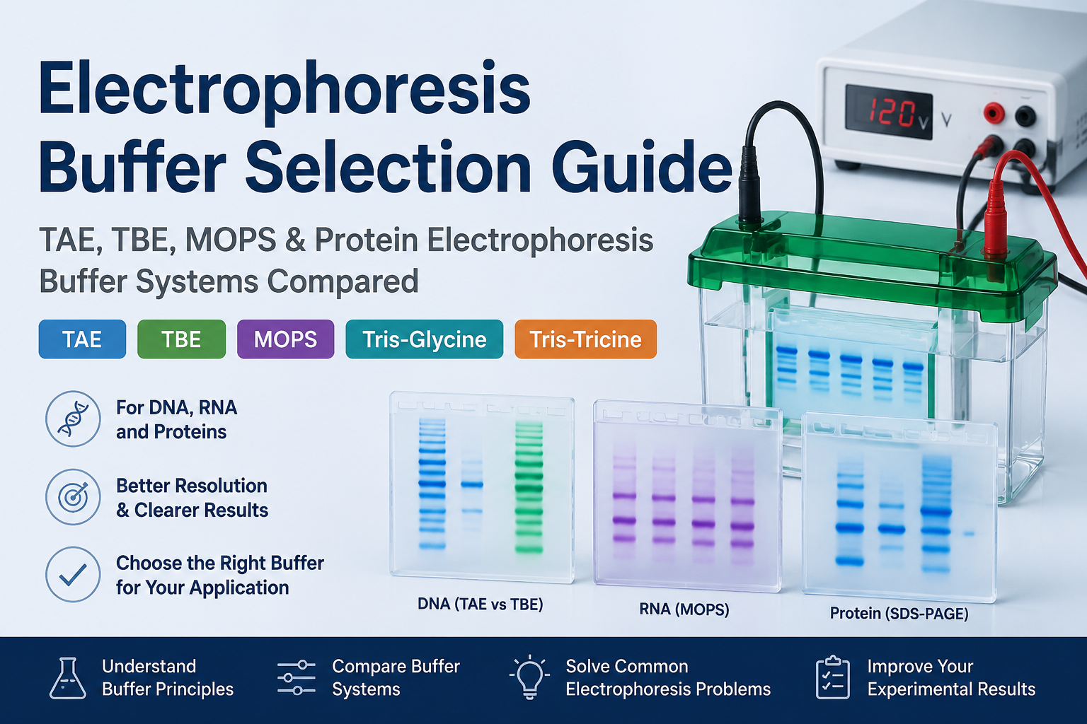
Introduction: Why Electrophoresis Buffer Selection Matters
Electrophoresis buffer plays a critical role in determining molecular migration speed, band resolution, heat generation, and downstream compatibility. Although DNA, RNA, and proteins can all be separated by electrophoresis, each type of molecule requires a different buffer system to achieve reliable and reproducible results.
The correct electrophoresis buffer should be selected based on:
· Sample type (DNA, RNA, or protein)
· Molecular size
· Gel type
· Native or denaturing conditions
· Need for sample recovery
· Downstream applications such as cloning, sequencing, or Western blotting 
Common electrophoresis buffer systems include:
· TAE and TBE buffers for DNA electrophoresis
· MOPS-based systems for RNA electrophoresis
· Tris-Glycine-SDS buffer for standard protein electrophoresis
· Tris-Tricine and Bis-Tris systems for specialized protein separation
Understanding the differences between these buffer systems helps researchers improve separation quality and avoid common electrophoresis problems.
1. How Electrophoresis Buffers Work
1.1 Maintaining pH and Molecular Charge
Electrophoresis depends on the movement of charged molecules through an electric field. The electrophoresis buffer maintains a stable pH environment to ensure consistent molecular charge and migration behavior.
For nucleic acids:
· DNA and RNA carry negative charges from their phosphate backbone.
· They migrate toward the positive electrode during electrophoresis.
For proteins:
· SDS binds to proteins and provides a relatively uniform negative charge.
· Protein separation in SDS-PAGE is mainly determined by molecular weight.
In contrast, Native PAGE preserves protein structure, meaning migration depends on:
· Molecular size
· Protein shape
· Surface charge
· Protein complex formation
1.2 Providing Conductivity and Controlling Heat Generation
Buffer ions provide electrical conductivity during electrophoresis.
An inappropriate ionic strength can negatively affect results:
· Low ionic strength
o Slow migration
o Poor band sharpness
o Increased diffusion
· High ionic strength
o Excessive current
o Increased heat generation
o Gel distortion
o Abnormal migration
Electrophoresis performance depends not only on buffer selection but also on:
· Buffer concentration
· Applied voltage
· Running time
· Gel thickness
1.3 Influence on Resolution and Downstream Applications
Different buffer systems have different:
· Buffer capacities
· Ionic compositions
· Conductivity levels
These factors influence:
· Band sharpness
· Migration speed
· Resolution
· Compatibility with downstream experiments
For example:
· TAE is commonly preferred for DNA recovery.
· TBE provides better small fragment resolution.
· MOPS is preferred for RNA denaturing electrophoresis.
· Tris-Glycine-SDS is widely used for routine protein analysis.
· Tris-Tricine improves separation of low molecular weight proteins.
2. DNA Electrophoresis Buffer Systems
2.1 TAE Buffer (Tris-Acetate-EDTA)
Composition and Principle
TAE buffer contains:
· Tris
· Acetic acid
· EDTA
Compared with TBE, TAE has lower ionic strength and generally allows faster DNA migration.
EDTA chelates divalent metal ions and helps reduce nuclease activity, improving nucleic acid stability.
Applications of TAE Buffer
TAE is commonly used for:
· PCR product analysis
· Restriction digestion analysis
· Plasmid verification
· Large DNA fragment separation
· DNA gel extraction
Because of its compatibility with many downstream enzymatic reactions, TAE is often preferred when DNA will be recovered from agarose gels for:
· Cloning
· Sequencing
· Ligation
· Restriction digestion
When Should You Choose TAE?
Choose TAE when your goal is:
✔ Routine DNA visualization
✔ DNA fragment recovery
✔ Downstream molecular biology applications
2.2 TBE Buffer (Tris-Borate-EDTA)
Composition and Principle
TBE contains:
· Tris
· Boric acid
· EDTA
It provides stronger buffering capacity than TAE and maintains stable pH during long electrophoresis runs.
TBE is particularly effective for separating small DNA fragments because it provides improved resolution.
Applications of TBE Buffer
TBE is commonly used for:
· Small DNA fragments
· Oligonucleotide analysis
· High-resolution DNA electrophoresis
· Nucleic acid PAGE
For fragments below approximately 100 bp or samples with very small size differences, TBE often provides clearer separation compared with TAE.
When Should You Choose TBE?
Choose TBE when your goal is:
✔ High-resolution DNA separation
✔ Small fragment analysis
✔ Long electrophoresis runs
2.3 SB Buffer (Sodium Borate Buffer)
SB buffer is a low ionic strength electrophoresis system that enables rapid DNA separation with reduced heat generation.
Advantages:
· Faster electrophoresis
· Lower heat production
Limitations:
· Less commonly used
· Lower compatibility with specialized applications
SB is suitable for:
· Quick PCR screening
· Simple DNA confirmation
For accurate fragment size analysis or DNA recovery, TAE or TBE is usually preferred.
2.4 RNA Electrophoresis Buffer Systems
MOPS Buffer for RNA Denaturing Electrophoresis
RNA differs from DNA because it easily forms secondary structures that can affect migration.
MOPS buffer is commonly used with:
· Formaldehyde agarose gels
· Denaturing RNA electrophoresis
It helps minimize RNA secondary structure effects and allows migration according to RNA size.
Applications include:
· Total RNA integrity analysis
· Northern blot preparation
· Large RNA fragment detection
RNA electrophoresis requires:
· RNase-free water
· RNase-free consumables
· Clean electrophoresis reagents
Urea-PAGE for Small RNA Analysis
Urea-PAGE provides high-resolution separation by:
· Using polyacrylamide gels
· Including urea to disrupt RNA secondary structures
Applications:
· miRNA analysis
· siRNA detection
· Small RNA separation
· Oligonucleotide analysis
3. Protein Electrophoresis Buffer Systems
3.1 Tris-Glycine-SDS Buffer System
Principle
Tris-Glycine-SDS is the most commonly used SDS-PAGE running buffer.
SDS denatures proteins and provides uniform negative charges, while the discontinuous buffer system enables:
1. Protein stacking in the stacking gel
2. Separation according to molecular weight in the resolving gel
Applications
Tris-Glycine-SDS is widely used for:
· Recombinant protein expression analysis
· Protein purification monitoring
· Western blot sample preparation
· Routine molecular weight determination
Limitations
For proteins below approximately 10 kDa:
· Bands may compress
· Resolution may decrease
· Small proteins may migrate close to the dye front
In these cases, alternative systems are recommended.
3.2 Tris-Tricine-SDS Buffer System
Tricine replaces glycine as the trailing ion, improving separation of low molecular weight proteins.
Applications:
· Small proteins
· Peptides
· Protein fragments
· Low molecular weight biomarkers
Advantages:
· Better resolution below 10 kDa
· Improved separation of small peptides
3.3 Bis-Tris/MES and Bis-Tris/MOPS Systems
Bis-Tris/MES
Suitable for:
· Low to medium molecular weight proteins
· High-resolution protein separation
Advantages:
· Sharp bands
· Mild operating conditions
Bis-Tris/MOPS
Suitable for:
· Medium to high molecular weight proteins
Advantages:
· Better migration of larger proteins
· Good reproducibility with pre-cast gels 
3.4 Native PAGE and Blue Native PAGE
Native PAGE
Unlike SDS-PAGE, Native PAGE does not denature proteins.
Protein migration depends on:
· Molecular weight
· Shape
· Charge
· Complex formation
Applications:
· Protein complexes
· Oligomerization studies
· Functional protein analysis
Blue Native PAGE
Blue Native PAGE uses Coomassie dye to provide proteins with negative charge while maintaining complex structures.
Applications:
· Membrane protein complexes
· Mitochondrial complexes
· Large protein assemblies
4. Electrophoresis Buffer Selection Guide
|
Experimental Goal |
Recommended Buffer System |
|
PCR product detection |
TAE or TBE |
|
DNA gel extraction |
TAE |
|
Small DNA fragment separation |
TBE |
|
RNA integrity analysis |
MOPS denaturing system |
|
Small RNA analysis |
Urea-PAGE |
|
Routine protein expression analysis |
Tris-Glycine-SDS |
|
Low molecular weight proteins |
Tris-Tricine-SDS |
|
Precast protein gels |
Bis-Tris/MES or Bis-Tris/MOPS |
|
Protein complex analysis |
Native PAGE / Blue Native PAGE |
5. Common Electrophoresis Problems and Solutions
5.1 Smiling Bands
Possible causes:
· Excessive voltage
· Buffer concentration too high
· Overheating
Solutions:
· Reduce voltage
· Replace old buffer
· Improve heat dissipation
5.2 Band Smearing
Possible causes:
DNA/RNA:
· Sample degradation
· Excess salt
· Overloading
Protein:
· Incomplete denaturation
· Protein degradation
· Buffer mismatch
Solutions:
· Improve sample quality
· Reduce loading amount
· Use the appropriate buffer system
5.3 Poor Resolution
Solutions:
· Small DNA fragments → Use TBE or PAGE
· Small proteins → Use Tris-Tricine or Bis-Tris/MES
· Large DNA fragments → Adjust agarose concentration
Buffer selection must always match:
· Sample size
· Gel concentration
· Experimental purpose
6. Frequently Asked Questions
TAE vs TBE: Which one should I use?
TAE is preferred for routine DNA electrophoresis and DNA recovery.
TBE is better for small fragments and high-resolution separation.
Quick guide:
· DNA recovery → TAE
· Small DNA fragments → TBE
· PCR confirmation → Either TAE or TBE
Can RNA electrophoresis use TAE or TBE?
No. For accurate RNA analysis, use MOPS denaturing systems or urea-PAGE because RNA secondary structures can affect migration.
Can SDS-PAGE running buffer be replaced with regular Tris buffer?
No. SDS-PAGE requires specific running buffer systems such as Tris-Glycine-SDS or Bis-Tris-based buffers.
How can I improve low molecular weight protein separation?
Use:
· Tris-Tricine-SDS buffer
· Bis-Tris/MES system
· Higher percentage resolving gels
Can electrophoresis buffer be reused?
Short-term reuse may be possible, but repeated use can change:
· Ionic composition
· Buffer capacity
· pH stability
For high-resolution experiments, RNA analysis, and quantitative comparisons, fresh buffer is recommended.
Conclusion: Choosing the Right Electrophoresis Buffer
Selecting the correct electrophoresis buffer is essential for achieving clear bands, accurate separation, and reproducible results.
General recommendations:
· DNA gel extraction → TAE
· Small DNA fragment analysis → TBE
· RNA integrity analysis → MOPS denaturing system
· Routine protein electrophoresis → Tris-Glycine-SDS
· Low molecular weight proteins → Tris-Tricine or Bis-Tris/MES
Matching the buffer system with the sample type, gel format, and experimental goal is the key to successful electrophoresis.


