High-quality cell suspensions are the foundation of reliable biological research. Whether the downstream application is single-cell RNA sequencing, flow cytometry, primary cell culture, cell sorting, immunology research, tumor microenvironment analysis, or drug screening, the quality of the starting cell suspension can directly determine the reliability of the final data.
After tissue dissociation, cell isolation, or sample processing, cell suspensions often contain more than just viable cells. They may also include dead cells, apoptotic cells, membrane fragments, red blood cells, organelle debris, protein aggregates, and other impurities. These contaminants are not always visible to the naked eye, but they can silently compromise experimental outcomes.
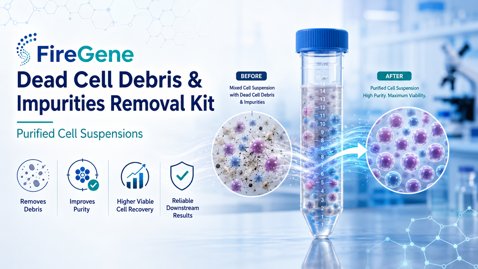
The FireGene Dead Cell Debris & Impurities Removal Kit is designed to help researchers obtain cleaner, more homogeneous, and more reliable cell suspensions by removing dead cell debris and unwanted impurities. This article explains why dead cell debris removal matters, how the kit supports purified cell suspension preparation, and how to troubleshoot common problems during density-based separation.
Why Dead Cell Debris Removal Matters
A dead cell debris removal kit is used to eliminate apoptotic cells, dead cells, cell fragments, and impurities from a cell suspension. The goal is to enrich viable cells and improve sample quality before downstream analysis.
Dead cells and debris are often underestimated. In many workflows, researchers focus on cell number and viability, but overlook small fragments and intracellular contaminants. However, dead cells release degraded RNA, enzymes, nucleases, cytoplasmic proteins, and intracellular metabolites into the suspension. These components can interfere with viable cells and distort downstream readouts.
For applications such as single-cell RNA sequencing, dead cell contamination can reduce RNA integrity, increase background signal, and affect cell capture efficiency. In flow cytometry, debris may interfere with gating accuracy and increase nonspecific background. In cell culture, dead cells and debris may promote stress responses, reduce proliferation, or increase contamination risk.
For this reason, removing dead cell debris is not simply a cleanup step. It is a critical sample preparation strategy that supports better data quality, reproducibility, and biological interpretation.
The Hidden Threat in Cell Suspensions
Dead cells and cellular debris can act as an invisible threat in experimental systems. Even when a suspension appears acceptable, microscopic debris and degraded intracellular contents may still be present.
These contaminants can affect experiments in several ways. First, they reduce the purity of viable cells in the final sample. Second, they may introduce degraded nucleic acids and proteins that interfere with molecular analysis. Third, they can increase viscosity or aggregation, making downstream processing less efficient. Finally, they may cause biased representation of cell populations, especially when fragile or rare cell types are lost during processing.
In advanced research workflows, poor sample quality is one of the most common bottlenecks. A purified cell suspension helps researchers reduce noise, protect viable cells, and generate more consistent results.
Designed for Advanced Cell-Based Research
The FireGene Dead Cell Debris & Impurities Removal Kit is particularly useful for workflows that require high-quality single-cell suspensions. These include:
Single-cell RNA sequencing: Clean suspensions improve cell capture, reduce debris-associated background, and support more accurate transcriptomic profiling.
Flow cytometry and FACS: Removing dead cells and impurities helps improve gating clarity, reduce background fluorescence, and support more reliable cell population analysis.
Primary cell culture: Cleaner suspensions reduce stress from dead cell components and help improve post-isolation cell recovery.
Tumor tissue research: Tumor-derived suspensions often contain necrotic debris and dead cells. Removing these impurities helps improve viable cell enrichment for tumor microenvironment studies.
Immune cell research: Spleen, blood-rich tissues, and inflammatory samples may contain red blood cells and debris that interfere with downstream immune profiling.
By improving cell suspension purity, the kit helps researchers obtain samples that are better suited for sensitive downstream applications.
How the Kit Works: Density-Based Separation
The FireGene Dead Cell Debris & Impurities Removal Kit uses a density-based separation workflow. In the procedure, a mixed solution containing the cell suspension and DRS① is carefully layered over DRS②. After centrifugation, different components separate into layers according to density.
The target viable cell fraction is generally collected from the buffy coat layer, while debris, dead cells, and heavier impurities are separated into other layers. Proper layering, reagent density, centrifuge settings, and careful aspiration are essential for successful separation.
Because the workflow depends on stable stratification, small operational changes can significantly affect the final result. The following troubleshooting guidance addresses common problems and optimization strategies.
How to Prevent Layer Infiltration During Sample Loading
One common issue is that the mixed solution quickly infiltrates into DRS② during layering, causing the two layers to merge. This prevents stable stratification and may reduce separation efficiency.
This problem is usually caused by an unstable density difference between DRS① and DRS② or by small-molecule impurities in the mixed solution. To avoid infiltration, DRS① and DRS② should be placed at 4°C for 30 minutes before use to ensure uniform reagent density.
Before layering, the mixed solution can be filtered once through a 20 μm cell sieve to remove small impurities such as fragmented organelles. During loading, researchers should use a 1 mL low-adhesion pipette tip and slowly add the mixed solution along the tube wall. Adding the solution drop by drop, with approximately two seconds between drops, helps each drop spread gently over the surface of DRS②. This improves stratification stability and supports cleaner layer formation.
What to Do When the Transition Layer Is Missing
After centrifugation, researchers should observe a clear multi-layer structure. If only three layers appear and the transition layer between DRS① and DRS② is missing, the centrifuge acceleration or deceleration may be too harsh. Rapid acceleration or braking can disrupt the reagent interface.
To solve this problem, set the centrifuge acceleration and deceleration to a medium setting. For digital centrifuges, this may correspond to a setting around “5,” depending on the instrument. Avoid sudden starts and stops.
Another practical method is to gently add 100 μL of PBS containing 2% FBS on top of DRS② before centrifugation. This creates a buffer layer and helps protect the interface. If needed, add an additional 0.5 mL DRS① to the mixed solution, then re-layer and centrifuge according to the procedure. These adjustments can help restore the expected four-layer structure and reduce dead cell contamination in the buffy coat layer.
Handling Tumor Samples With High Dead Cell Rates
Tumor-derived single-cell suspensions often contain large amounts of necrotic debris, especially when the dead cell rate exceeds 40%. After centrifugation, the debris layer may become very thick and cover the buffy coat layer, making it difficult to identify viable cells.
In this situation, use a stepwise aspiration and microscopic observation strategy. Slowly aspirate the upper debris layer with a 1 mL low-adhesion pipette tip. After removing approximately 200 μL each time, take 10 μL of the aspirated liquid and observe it under a microscope.
When uniformly distributed round viable cells begin to appear, stop removing the debris layer. The buffy coat layer is usually located about 2–3 mm below the liquid surface at this point. Insert the pipette tip carefully along the tube wall and slowly aspirate approximately 800 μL of liquid. If the collected fraction still contains debris, filter it again through a 20 μm cell sieve. With careful handling, viable cell purity can reach more than 90%.
How to Obtain a Dense Cell Pellet
After collecting the buffy coat layer, the sample is typically diluted with PBS containing 5% FBS and centrifuged. Sometimes the resulting cell pellet may appear loose and flocculent, and cells may be lost when the supernatant is discarded.
This problem can occur because viable cells carry a negative surface charge, causing mutual repulsion and poor aggregation. To improve pellet density, centrifugation can be adjusted to 500 × g for 8 minutes. During dilution, adding 30 μL of 1% polylysine to 10 mL PBS can help neutralize surface charge and improve cell aggregation.
After centrifugation, do not remove all supernatant aggressively. Retain approximately 150 μL of supernatant and gently pipette to resuspend the pellet. This reduces the risk of losing small cell pellets and improves recovery.
Reagent Storage and Validity
Storage conditions are important for maintaining kit performance. DRS① should be stored at 4°C in the dark and used within its recommended validity period. If DRS① has been stored for more than one year after opening, its density-regulating components may degrade.
A practical verification test can be performed by mixing DRS① with RPMI 1640 containing 2% FBS, layering it over DRS②, and centrifuging under the recommended conditions. If the buffy coat volume is too small and the dead cell proportion remains high, the reagent may be invalid. Expired or partially degraded reagent is not recommended for sensitive applications such as single-cell sequencing.
Managing Red Blood Cell Interference
Spleen samples and blood-rich tissues may contain many red blood cells. Because red blood cells can have a density similar to viable cells, the boundary between the buffy coat layer and red blood cell layer may become blurred.
After centrifugation, place the tube on ice and allow it to stand for 10 minutes. Red blood cells will slowly settle because of their slightly higher density, helping form a clearer interface. Researchers can then use a small-volume low-adhesion pipette tip to sample and verify the layer under a microscope. Aspirating only 600–800 μL from the correct region helps reduce red blood cell contamination.
Avoiding Cell Adhesion During Resuspension
Cell adhesion to the tube wall can reduce the actual cell concentration in the mixed solution. To prevent this, pre-coat the centrifuge tube with RPMI 1640 containing 2% FBS for 5 minutes before use. This forms a simple anti-adhesion layer on the tube wall.
During resuspension, use a low-adhesion pipette tip and pipette gently along the tube wall. Avoid direct, forceful pipetting that may drive cells onto the plastic surface. If cells have already adhered, gently collect them with a sterile cell scraper. These steps can improve overall cell recovery.
Using DMEM Instead of RPMI 1640
RPMI 1640 is generally preferred for resuspension. If DMEM is used instead, density differences may affect stratification and reduce viable cell recovery. DMEM has a higher density than RPMI 1640, especially when serum is added.
To improve performance with DMEM, use serum-free DMEM, increase the cell concentration to approximately 2 × 10⁶ cells/mL, and slightly reduce centrifugation speed from 1400 × g to 1350 × g. These adjustments can help restore separation performance and improve viable cell recovery.
Conclusion
The FireGene Dead Cell Debris & Impurities Removal Kit provides an optimized solution for preparing purified cell suspensions by removing dead cells, apoptotic fragments, debris, and impurities. For modern research workflows, especially single-cell sequencing, flow cytometry, primary culture, and tumor microenvironment analysis, sample quality is not optional. It is the foundation of reliable data.
By following proper layering techniques, maintaining reagent density, controlling centrifugation conditions, carefully collecting the buffy coat layer, and minimizing cell loss, researchers can improve viable cell purity, recovery rate, and downstream reproducibility.
Clean cells lead to clearer data. For researchers working with complex or fragile cell samples, effective dead cell debris removal can make the difference between noisy results and reliable biological insight.
FireGene, light your research with passion, innovation, and profession.
FireGene Single-Cell Sample Prep
Looking for validated dissociation kits?
FireGene's tissue dissociation kits are optimized for specific organ types — brain, tumor, liver, GI, reproductive, and more. Validated for 10x Genomics Chromium and BD Rhapsody workflows.


