Preparing high-quality single-cell suspensions from lung tissue is one of the most technically demanding steps in single-cell RNA sequencing workflows. Unlike many solid tissues, lung samples contain complex alveolar architecture, abundant extracellular matrix, vascular networks, epithelial barriers, mucus-producing cells, and, in disease models, inflammatory debris or excess red blood cells. These features can make tissue dissociation inconsistent, reduce cell yield, compromise cell viability, and introduce impurities that interfere with downstream scRNA-seq library preparation.
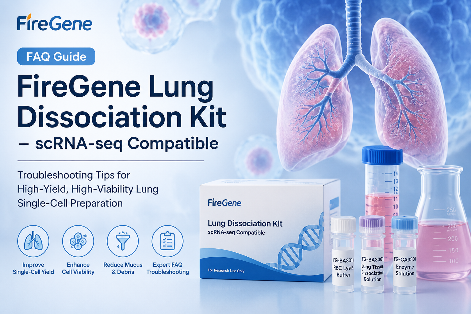
The FireGene Lung Dissociation Kit – scRNA-seq Compatible is designed to support lung tissue processing for single-cell applications, but successful use still depends on precise sample preparation, optimized enzymatic digestion, and careful handling of fragile pulmonary cell populations. This FAQ-based guide summarizes common problems researchers may encounter during lung tissue dissociation and provides practical troubleshooting strategies to help improve cell yield, viability, filtration efficiency, and sample quality. The guidance below is based on the provided FireGene FAQ material.
Why does lung tissue sometimes remain as intact alveoli after enzymatic digestion?
One common issue during lung dissociation is that the alveolar structure remains largely intact even after extended enzymatic digestion. Researchers may observe many complete alveoli after one hour of enzymolysis, resulting in a very low single-cell yield.
The primary reason is usually insufficient tissue mincing. Lung tissue has a highly elastic and air-filled architecture. If the tissue pieces are too large, the alveolar cavities may not be fully exposed. As a result, the enzymatic solution cannot efficiently contact the alveolar wall fibers, and the tissue remains structurally intact.
To improve alveolar disruption, mince the lung tissue into pieces smaller than 0.2 mm³, approximately one-third the size typically used for regular tissue processing. This helps rupture the alveolar cavities and exposes more tissue surface area to the dissociation solution. In addition, repeatedly puncturing the minced tissue with a 1 mL syringe needle before enzymatic digestion can physically disrupt the alveolar walls and improve enzyme penetration.
For particularly resistant samples, an additional 50 μL of lung tissue dissociation solution may be added during enzymolysis, and the digestion time can be extended to 1.5 hours. According to the FAQ guidance, this adjustment can increase alveolar structure destruction to more than 85% and improve single-cell yield by approximately 60%.
How can viscous mucus be removed from COPD lung tissue samples?
Lung tissue from chronic obstructive pulmonary disease models often contains excessive mucus. During dissociation, this mucus may mix with enzymolysis products, wrap around cells, and clog filtration membranes. This can significantly slow filtration and reduce recovery of usable single cells.
The mucus is mainly caused by mucin secreted by COPD lung tissue, combined with digestion products released during enzymatic processing. To reduce viscosity, add 100 μL of 0.1% hyaluronidase during enzymolysis. Hyaluronidase helps degrade mucopolysaccharide components in the mucus and improves suspension fluidity.
Before filtration, centrifuge the cell suspension at 4°C, 500 × g for 3 minutes, then discard the upper mucus layer. This step helps separate viscous material from the cell-containing fraction. During filtration, rinse the 70 μm cell strainer three times with PBS containing 2% FBS to help mucus pass through the filter and prevent clogging. These adjustments can increase filtration speed by approximately 50%, improving sample processing efficiency for mucus-rich lung tissues.
What causes liquid stratification during hybridization oven digestion?
When using a hybridization oven for enzymatic digestion, researchers may notice that the liquid separates into a clear upper layer and a turbid lower layer. This stratification leads to uneven digestion because tissue fragments are not consistently exposed to the enzymatic solution.
The cause is generally related to the density and composition of lung tissue. Lung samples contain blood vessels, fibrous material, and extracellular components that can settle at the bottom of the centrifuge tube if rotation is insufficient. When mixing is inadequate, enzyme contact becomes uneven, and some tissue fragments remain under-digested.
To solve this, increase the hybridization oven rotation speed from 20–30 rpm to 25–35 rpm to enhance liquid convection. For better mixing, place one sterile 3 mm magnetic stir bar into the centrifuge tube before enzymolysis. Manual intervention can also help: invert the centrifuge tube once every 15 minutes to ensure more uniform contact between tissue and dissociation solution. According to the FAQ, these changes can improve enzymatic digestion uniformity by approximately 70%.
How can researchers check whether the dissociation solution has lost enzyme activity?
After opening the kit, the lung tissue dissociation solution may lose activity if stored for too long or handled improperly. If the solution has been stored at -20°C for five months and tissue remains in blocks after digestion, enzyme activity may have declined significantly.
A quick way to evaluate the solution is to perform a mini enzymolysis experiment. Mix 50 μL of dissociation solution with 10 mg of minced lung tissue, then incubate at 37°C for 30 minutes. After incubation, pipette the sample 10 times. If the tissue pieces remain larger than 1 mm³ and no obvious cells are released, the dissociation solution should be considered completely inactive.
If only a small number of cells are released but digestion is insufficient, the amount of dissociation solution can be doubled to 480 μL, and the enzymolysis time can be extended to 2.5 hours. However, this workaround is only suitable for basic experiments and is not recommended for single-cell sequencing, where high viability and reproducibility are essential.
How should newborn mouse lung tissue be dissociated without excessive cell death?
Newborn mouse lung tissue, especially from mice within three days of birth, is highly fragile. Researchers may find that cell viability drops to around 40% after standard dissociation.
This happens because immature lung cells have delicate cell membranes that are easily damaged by enzymatic digestion and mechanical force. To protect these cells, reduce the enzymolysis time from the standard range of 0.5–2 hours to only 20–30 minutes. Use a water bath rather than aggressive shaking equipment, and gently shake the sample every 5 minutes.
For washing, replace regular PBS containing 5% FBS with PBS containing 10% FBS. The higher serum concentration provides better cell protection during centrifugation and washing. With these adjustments, cell viability can be increased to more than 75%, making the suspension more suitable for downstream single-cell analysis.
How can black impurity particles be removed from the cell pellet?
After lung tissue dissociation and centrifugation, black particles may appear in the cell pellet. These particles may be carbon dust inhaled into lung tissue or aggregates formed by cell debris. They can interfere with cell counting, staining, microscopy, and downstream data quality.
To remove them, gently resuspend the pellet in 1 mL of PBS containing 5% FBS. Let the suspension stand for 2 minutes so that the heavier black particles settle. Carefully aspirate the upper cell suspension and transfer it into a new centrifuge tube. Repeat this procedure twice.
This simple sedimentation-based cleanup can remove more than 80% of black particles while keeping cell viability loss at or below 5%. It is especially useful for lung samples from animals exposed to environmental particulates, smoke, or inflammatory injury models.
What can be done after accidental over-digestion?
Accidental over-digestion can occur when enzymolysis is extended unintentionally, for example to 3 hours in a water bath. In this case, cell viability may drop to around 30%, making the sample unsuitable for sensitive applications such as scRNA-seq.
Although over-digestion cannot be fully reversed, some viable cells may be recovered by gradient-style centrifugation. First, centrifuge the cell suspension at 4°C, 200 × g for 3 minutes, then discard the upper layer containing many dead cells and debris. Next, resuspend the pellet in 5 mL of PBS containing 10% FBS and centrifuge again at 4°C, 300 × g for 5 minutes.
After resuspension, stain with trypan blue to evaluate viability. Viable cells are usually concentrated in the lower portion of the pellet. This method may recover cells with viability of at least 60%, but the yield may be only about 30% of the original. Therefore, rescued samples are more appropriate for low-cell-number applications such as cell culture, rather than high-quality single-cell sequencing.
How can red blood cell lysis be optimized to protect pulmonary endothelial cells?
Lung tissue is highly vascularized, so red blood cell contamination is common. In some preparations, red blood cells may account for more than 50% of the cell suspension. However, treatment with red blood cell lysis buffer can damage sensitive pulmonary vascular endothelial cells, reducing viability from 80% to 50%.
To reduce toxicity, dilute the FG-BA3311 Red Blood Cell Lysis Buffer one-fold with PBS. Perform lysis at 4°C and shorten the incubation time to 2–3 minutes. Monitor the reaction under a microscope and stop the lysis immediately once red blood cells are visibly lysed.
After lysis, wash the cells three times with PBS containing 10% FBS to remove residual lysis buffer. This optimized protocol can help maintain pulmonary vascular endothelial cell viability above 70%, improving preservation of vascular cell populations for single-cell analysis.
Best Practices for Lung Single-Cell Dissociation
For scRNA-seq-compatible lung dissociation, the key goals are high cell yield, high viability, minimal debris, and preservation of diverse cell populations. Based on the FAQ guidance, several practical principles emerge.
First, mechanical preparation matters. Lung tissue must be minced more finely than many other tissues because intact alveoli can resist enzymatic access. Second, disease-model samples often require customized handling. COPD lung tissue may need mucus-reduction steps, while vascularized or inflamed tissue may require carefully optimized red blood cell lysis. Third, cell type sensitivity should guide protocol adjustments. Newborn lung cells and pulmonary endothelial cells require gentler digestion, lower temperature exposure, shorter incubation, and enhanced serum protection.
Finally, quality control should be performed before committing samples to single-cell sequencing. Assess digestion completeness, cell viability, red blood cell contamination, debris levels, and filtration performance. If enzyme activity is questionable, a mini enzymolysis test can prevent wasting valuable samples on suboptimal reagents.
Conclusion
The FireGene Lung Dissociation Kit – scRNA-seq Compatible supports preparation of lung single-cell suspensions for downstream single-cell workflows, but optimal results depend on tissue-specific troubleshooting. Problems such as intact alveoli, mucus-rich suspensions, liquid stratification, reduced enzyme activity, fragile neonatal cells, impurity particles, over-digestion, and red blood cell contamination can all affect sample quality.
By adjusting mincing size, digestion time, enzyme volume, mixing conditions, washing buffers, centrifugation parameters, and red blood cell lysis conditions, researchers can improve single-cell yield and viability while preserving key pulmonary cell populations. For lung scRNA-seq studies, these details are not minor protocol refinements—they are critical determinants of data quality, cellular representation, and experimental reproducibility.
FireGene, light your research with passion, innovation, and profession.
FireGene Single-Cell Sample Prep
Looking for validated dissociation kits?
FireGene's tissue dissociation kits are optimized for specific organ types — brain, tumor, liver, GI, reproductive, and more. Validated for 10x Genomics Chromium and BD Rhapsody workflows.


