In tumor microenvironment research, high-quality single-cell suspension is one of the most critical starting points for downstream analysis. Whether researchers are preparing samples for flow cytometry, single-cell sequencing, primary tumor cell culture, immune profiling, or cell sorting, the quality of tumor tissue dissociation directly affects cell yield, cell viability, and data reliability.
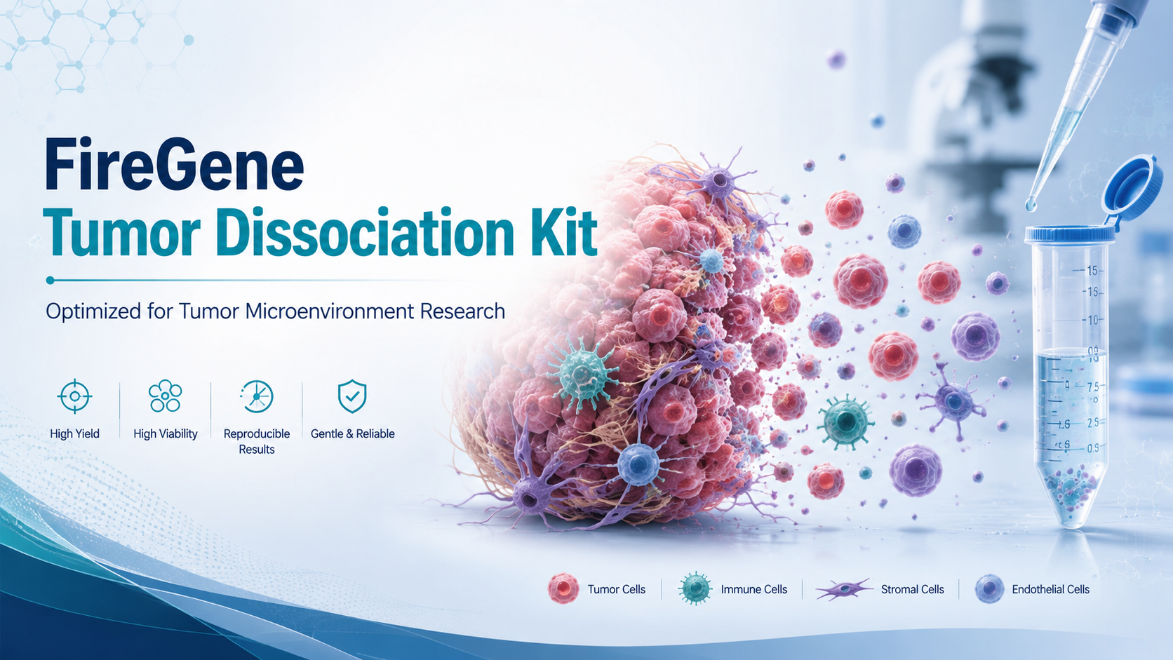
The FireGene Tumor Dissociation Kit is designed to support efficient dissociation of fresh solid tumor tissues while preserving viable cells for downstream tumor microenvironment studies. Tumor tissues are often structurally complex, containing malignant cells, stromal cells, immune cells, extracellular matrix components, collagen fibers, necrotic areas, and red blood cells. A dissociation workflow must therefore be strong enough to break down tissue architecture but gentle enough to maintain cell integrity.
This article answers key practical questions about using the FireGene Tumor Dissociation Kit and explains how to optimize tumor tissue preparation for reliable single-cell suspension generation.
What Is the FireGene Tumor Dissociation Kit Used For?
The FireGene Tumor Dissociation Kit is mainly intended for fresh solid tumor tissues, including commonly studied tumor types such as lung cancer, breast cancer, and liver cancer tissues. These samples typically require enzymatic digestion to break down extracellular matrix and tissue structure, allowing researchers to obtain a usable single-cell suspension.
The kit is especially suitable for tumor microenvironment workflows because it helps release diverse cellular components from solid tumors. These may include tumor cells, immune infiltrates, fibroblasts, endothelial cells, and other stromal populations. By supporting controlled dissociation, the kit can help researchers improve sample consistency before downstream analysis.
However, the kit is not suitable for non-solid tumor samples, such as leukemia or lymphoma cell suspensions. Hematological tumor cells are usually already in suspension and do not require tissue dissociation. Using a solid-tissue enzymatic system on suspended tumor cells may cause severe cell damage or massive cell death. For these samples, a dedicated cell sorting or suspension-cell processing kit is more appropriate.
Can Cryopreserved Tumor Tissue Be Used?
Fresh tumor tissue is strongly preferred. Cryopreserved and resuscitated tumor tissues may be used only with caution. The freezing and thawing process can damage tumor tissue structure and reduce cell viability after dissociation. Compared with fresh tissues, cryopreserved tumor samples may show a 20%–30% decrease in cell viability.
Before using a thawed tumor sample, researchers should perform a pre-quality inspection with trypan blue staining. A sample viability of at least 60% is recommended before proceeding. If viability is lower than this threshold, the tissue may not produce a reliable single-cell suspension and may be unsuitable for sensitive downstream applications such as single-cell RNA sequencing.
Why Tissue Weight Matters
The recommended tissue input is approximately 200 mg of fresh tumor tissue. Tissue quantity plays a major role in dissociation performance because the ratio between tissue mass and enzymatic dissociation solution determines digestion efficiency.
If the tissue amount is too low, such as 80 mg, the dissociation solution becomes relatively excessive. This may expose cells to a high enzyme-to-tissue ratio, increasing the risk of over-digestion and leading to a significant decrease in cell viability. In some cases, viability may drop by more than 40%.
For insufficient tissue input, it is better to combine multiple small tissue pieces until the total reaches approximately 180–200 mg, then use the standard reagent volume.
If the tissue amount is too high, such as 300 mg, dissociation may also be compromised. A 5 mL centrifuge tube may not provide enough space for proper tissue dispersion. Crowded tissue fragments can prevent uniform enzymatic exposure, resulting in incomplete digestion and excessive tissue debris.
For larger tissue samples, the tissue should be divided into two tubes. Each tube should contain approximately 180–220 mg of tissue with the corresponding dissociation solution and medium volume. This helps maintain the correct enzyme-to-tissue ratio and improves enzymatic efficiency.
How to Choose the Right Digestion Time
The recommended digestion window is 0.5–2 hours, but the optimal starting time depends on tissue density and tumor type.
Low-density tumor tissues, such as some lung cancer tissues or relatively loose tumor lesions, usually require a shorter digestion time of 0.5–1 hour. These tissues often contain less fibrotic material and are easier to dissociate.
High-density tumor tissues, such as breast cancer and liver cancer tissues, generally require a longer initial digestion time of 1–1.5 hours. These tissues often contain more collagen fibers and extracellular matrix components, requiring more time for enzymatic decomposition.
Both under-digestion and over-digestion can affect results. If digestion is insufficient, the tissue may remain in large clusters, with many cell aggregates containing more than 10 cells. This can reduce single-cell yield to below 50%.
If digestion is excessive, tumor cells may become damaged, viability may fall below 50%, and excessive cell debris may interfere with downstream single-cell sequencing or cell analysis. For best results, quality inspection should be performed every 20 minutes during digestion to determine the appropriate termination time.
Why 70 μm Filtration Is Important
Filtration is a key step in preparing a clean single-cell suspension. The protocol recommends using a 70 μm cell strainer and rinsing the centrifuge tube three times to collect a total of 12 mL filtrate.
Skipping one rinse can cause a substantial loss of residual tumor cells. Tumor cells may adhere to the inner wall of the centrifuge tube, and each rinse contributes to cell recovery. Omitting one rinse may reduce final cell yield by approximately 25%–30%.
The 70 μm pore size is also important. A 50 μm strainer is not recommended because it may clog easily with fiber debris and increase mechanical stress on cells. Although some small tumor cells can physically pass through the pores, blockage can cause filtration difficulty and reduce viability.
A 100 μm strainer is also unsuitable because it may allow undigested tissue fragments to pass into the suspension. These fragments can interfere with cell counting and may block microfluidic chips used in single-cell sequencing. Therefore, 70 μm filtration offers a practical balance between removing debris and preserving cell recovery.
Can DMEM Replace RPMI 1640?
Yes. DMEM can replace RPMI 1640 medium in the dissociation workflow. After substitution, there is no need to adjust centrifugation conditions or digestion time. The centrifugation parameters remain 4°C, 300 × g for 5 minutes.
Both DMEM and RPMI 1640 are commonly used basal media for mammalian cells. Although they differ in glucose and amino acid composition, both can maintain suitable osmotic pressure and pH during tumor tissue dissociation. According to the FAQ data, breast cancer tissue dissociation using these two media showed a cell viability difference of 5% or less.
Researchers may therefore choose the medium based on laboratory availability. If the dissociated cells will be used for subsequent culture, it is preferable to use the same medium as the downstream culture system to reduce cellular adaptation stress.
When Should Red Blood Cell Lysis Be Performed?
Some tumor tissues, especially blood-rich tumors, may contain many red blood cells. If red blood cell removal is necessary, BA3311 Red Blood Cell Lysis Buffer can be used after the washing steps and before final resuspension.
The recommended workflow is to perform red blood cell lysis after the second wash. Add 1 mL of lysis buffer, incubate at 4°C for 5 minutes, then centrifuge at 4°C, 300 × g for 5 minutes. After removing the supernatant, resuspend the cells with PBS containing 5% FBS and perform an additional wash before proceeding.
Temperature and timing are important. Lysis should not exceed 8 minutes, and incubation at 4°C helps reduce potential damage to tumor cells. If red blood cells are abundant, lysis may be repeated once, but an additional PBS wash is necessary to remove residual lysis buffer and protect downstream applications.
How Should the Dissociation Reagent Be Stored?
The tumor tissue dissociation solution should be stored at -20°C and has a validity period of two years. If the ice pack melts during shipping and the reagent remains at 4°C for up to 3 hours, it can still be used. However, it should be returned to -20°C immediately and fully mixed before use.
Repeated freeze-thaw cycles should be avoided. Each freeze-thaw cycle may reduce enzyme activity by 12%–18%. After more than three freeze-thaw cycles, enzyme activity may fall below 50%, which can lead to incomplete extracellular matrix digestion and poor single-cell suspension quality.
To prevent activity loss, the dissociation solution can be aliquoted into single-use volumes after receipt. For example, 250 μL aliquots can be stored at -20°C, with one aliquot used per experiment.
What If Tumor Cells Remain in the Supernatant?
After centrifugation, if many suspended tumor cells are still visible in the supernatant, the supernatant should not be discarded immediately. Instead, collect it and re-centrifuge at 4°C, 350 × g for 8 minutes. This slightly increased speed and extended time can improve cell recovery.
However, centrifugation should not be made too harsh. Speeds above 400 × g or times longer than 10 minutes are not recommended because excessive force may damage tumor cells and reduce viability.
Can the Final Cell Suspension Be Stored?
The final tumor cell suspension should ideally be used immediately. If immediate downstream processing is not possible, short-term storage is allowed at 4°C for no more than 1 hour.
The cell concentration should be adjusted to 1 × 10⁶–1 × 10⁷ cells/mL using PBS containing 5% FBS, and the suspension should be kept in a low-adhesion centrifuge tube. Repeated shaking should be avoided.
After one hour, tumor cell viability may decrease by 10%–12% per hour, and cell aggregation becomes more likely. If stored for more than three hours, viability may fall below 50%, making the sample unsuitable for single-cell sequencing or cell culture.
Before use, the sample should be checked again. A viability of at least 65% and a single-cell ratio of at least 75% are recommended for downstream experiments.
Conclusion
The FireGene Tumor Dissociation Kit provides a practical workflow for preparing single-cell suspensions from fresh solid tumor tissues. By controlling tissue input, digestion time, filtration conditions, washing steps, centrifugation parameters, and storage time, researchers can improve cell yield and viability while reducing debris and aggregation.
For tumor microenvironment research, sample preparation quality is directly linked to downstream data quality. A well-optimized dissociation protocol helps preserve biologically relevant cell populations and supports more reliable analysis in applications such as single-cell sequencing, flow cytometry, cell sorting, and tumor cell culture.
FireGene, light your research with passion, innovation, and profession.
FireGene Single-Cell Sample Prep
Looking for validated dissociation kits?
FireGene's tissue dissociation kits are optimized for specific organ types — brain, tumor, liver, GI, reproductive, and more. Validated for 10x Genomics Chromium and BD Rhapsody workflows.


