
Introduction
Single-cell analysis has become an important approach for investigating the cellular complexity of the central nervous system. Among neural tissues, the spinal cord presents particular technical challenges because of its dense extracellular matrix, extensive neuronal projections, abundant glial populations, and sensitivity to mechanical stress. Efficient preparation of a high-quality single-cell suspension is therefore a critical step for downstream applications such as single-cell RNA sequencing, flow cytometry, cell sorting, primary cell culture, and molecular profiling.
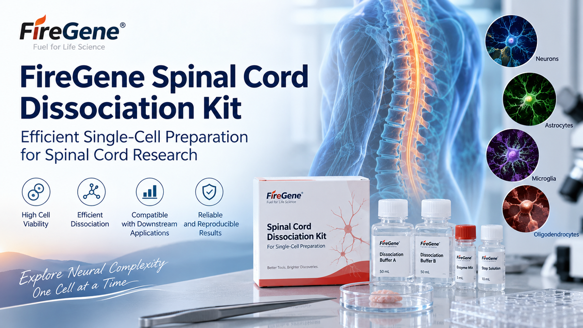
The FireGene Spinal Cord Dissociation Kit is designed to support the preparation of single-cell suspensions from spinal cord tissue under controlled laboratory conditions. By combining optimized enzymatic dissociation with standardized handling procedures, the kit helps researchers reduce excessive mechanical damage while improving cell recovery and suspension quality.
Reliable tissue dissociation is particularly important in spinal cord research because inappropriate digestion conditions can lead to reduced cell viability, selective loss of vulnerable cell populations, increased debris, and changes in cellular gene-expression profiles. A standardized dissociation workflow can therefore improve the reproducibility of experiments involving neural and glial cell populations.
Why Spinal Cord Tissue Is Difficult to Dissociate
Spinal cord tissue contains a highly organized network of neurons, astrocytes, oligodendrocytes, microglia, endothelial cells, pericytes, immune cells, and other specialized cell types. These cells are embedded within extracellular matrix components and interconnected through complex cellular processes.
Unlike many soft tissues, spinal cord samples can be difficult to convert into homogeneous single-cell suspensions without compromising cell integrity.
Several factors contribute to this difficulty.
First, neuronal cells possess long axons and dendritic processes that can be easily damaged during aggressive mechanical dissociation. Excessive pipetting, grinding, or vortexing may cause membrane disruption and increased release of intracellular components.
Second, neural tissues contain substantial lipid-rich material, particularly because of myelin. During tissue dissociation, myelin fragments can contribute to debris formation and may interfere with cell counting, flow cytometry, cell sorting, or microfluidic single-cell workflows.
Third, extracellular matrix proteins and cell-cell adhesion structures can limit the efficiency of purely mechanical dissociation. Controlled enzymatic digestion is therefore commonly required to release individual cells from spinal cord tissue.
Finally, different cellular populations have different sensitivities to enzymatic exposure. Excessive digestion may reduce surface-marker integrity or negatively affect cell viability, whereas insufficient digestion can result in large aggregates and poor cell recovery.
For these reasons, spinal cord dissociation requires a balance between effective tissue digestion and preservation of cellular quality.
Principle of Enzymatic Spinal Cord Dissociation
The FireGene Spinal Cord Dissociation Kit uses an enzyme-based tissue-processing strategy designed to disrupt extracellular and intercellular structures while minimizing unnecessary cellular damage.
A typical spinal cord dissociation workflow can be summarized as:
Spinal Cord Collection → Tissue Mincing → Enzymatic Digestion → Gentle Mechanical Dissociation → Filtration → Washing → Single-Cell Suspension

During the enzymatic digestion stage, tissue-associated proteins and extracellular matrix components are progressively disrupted. This facilitates the release of individual cells and reduces the amount of mechanical force required during later processing.
The combination of enzymatic digestion and gentle trituration is particularly useful for neural tissues. Enzymes loosen the structural organization of the tissue, while controlled pipetting helps separate remaining cell clusters.
The objective is not simply to maximize the total number of released cells. Instead, an effective dissociation protocol should generate a suspension with several characteristics:
high cell viability;
low levels of cellular debris;
minimal cell aggregation;
reproducible cell yield;
preservation of major cellular populations;
and compatibility with downstream analytical methods.
These parameters are particularly important in single-cell experiments, where poor starting material can strongly influence data quality.
Importance of Cell Viability in Single-Cell Preparation
Cell viability is one of the most important quality indicators following spinal cord tissue dissociation.
Dead or damaged cells can release nucleic acids, proteins, and cellular fragments into the suspension. Extracellular DNA may increase sample viscosity and promote cell aggregation, while membrane fragments and other debris can interfere with accurate cell counting.
For single-cell RNA sequencing, excessive numbers of damaged cells may contribute to increased ambient RNA and background signals. This can complicate cell-type identification and gene-expression analysis.
Similarly, for flow cytometry and fluorescence-activated cell sorting, poor-quality suspensions may produce abnormal scatter profiles and increase the proportion of unwanted events.
Several experimental variables can influence viability during spinal cord dissociation, including:
Tissue Processing Time
Spinal cord tissue should generally be processed efficiently after collection. Prolonged delays may increase cellular stress and reduce recovery of sensitive populations.
Tissue Fragment Size
Before enzymatic digestion, the tissue is typically cut into small pieces. Appropriate fragmentation increases the surface area available for enzyme exposure and can improve digestion efficiency.
Digestion Duration
Insufficient digestion may leave large tissue fragments, whereas prolonged digestion can damage cell membranes or alter surface proteins. Researchers should carefully control incubation time according to tissue quantity and experimental requirements.
Mechanical Force
Excessive mechanical force is a common cause of reduced cell viability. Gentle and progressive trituration is generally preferable to vigorous pipetting or homogenization.
Temperature Control
Temperature should be managed throughout sample preparation according to the protocol because enzymatic activity and cellular metabolism are strongly temperature dependent.
Standardizing these variables helps reduce experiment-to-experiment variability.
Reducing Debris and Cell Aggregation
Debris is a frequent challenge in spinal cord single-cell preparation.
Neural tissue contains myelin, extracellular matrix fragments, disrupted cellular processes, and membrane components. These materials can remain in the suspension after digestion and may negatively affect downstream analysis.
After enzymatic dissociation, filtration is commonly used to remove undigested tissue fragments and larger aggregates. Appropriate cell strainers can help produce a more uniform suspension suitable for counting and further processing.
Cell aggregation should also be monitored carefully. Aggregates can interfere with cell concentration measurements and may result in doublets or multiplets in single-cell sequencing workflows.
Gentle resuspension and appropriate washing can help reduce aggregation. When necessary, researchers may also incorporate additional debris-removal or dead-cell-removal procedures depending on tissue condition and downstream application.
However, every additional purification step may result in some loss of viable cells. Therefore, the degree of purification should be selected according to the experimental objective.
For example, a highly purified suspension may be particularly important for droplet-based single-cell RNA sequencing, whereas some cell culture applications may tolerate a slightly higher level of debris.
Applications in Single-Cell Research
High-quality spinal cord cell suspensions can support a broad range of experimental applications.
Single-Cell RNA Sequencing
Single-cell RNA sequencing allows researchers to investigate transcriptional heterogeneity among individual cells. In spinal cord research, this approach is used to characterize neuronal and glial populations, examine cellular states, and study changes in gene expression under different experimental conditions.
The quality of the initial cell suspension has a major influence on sequencing performance. Cell concentration, viability, aggregation, and debris levels should therefore be evaluated before library preparation.
Flow Cytometry
Flow cytometry can be used to characterize specific spinal cord cell populations using fluorescent markers.
Researchers may investigate microglial populations, immune-cell infiltration, glial markers, endothelial populations, or other cellular subsets. Reliable dissociation helps ensure that cells pass through the flow cytometer as individual events rather than aggregates.
Fluorescence-Activated Cell Sorting
Fluorescence-activated cell sorting enables researchers to isolate selected cell populations for downstream molecular analysis or culture.
A well-dissociated sample can improve sorting efficiency and reduce clogging of the instrument.
Primary Cell Research
Spinal cord-derived cells may also be used for primary culture or short-term experimental studies. Because many neural cell populations are sensitive to processing stress, gentle dissociation can be important for maintaining cellular function after isolation.
Molecular and Cellular Profiling
Prepared cell suspensions may support downstream assays involving RNA extraction, protein analysis, receptor expression, signaling pathways, inflammatory responses, and other cellular measurements.
Experimental Considerations for Reproducible Results
Reproducibility is essential in spinal cord dissociation experiments.
Researchers should standardize major experimental variables wherever possible, including tissue mass, animal age, anatomical sampling region, digestion time, enzyme concentration, temperature, and mechanical dissociation method.
The ratio between tissue quantity and digestion solution is particularly important. Processing excessive tissue in an insufficient volume of digestion reagent may lead to incomplete dissociation.
Researchers should also avoid overloading filtration devices because excessive tissue material can reduce filtration efficiency.
Before beginning downstream experiments, several quality-control parameters can be assessed:
Cell concentration: Determine whether sufficient cells have been recovered for the intended assay.
Cell viability: Evaluate the proportion of viable cells using an appropriate viability method.
Suspension uniformity: Examine whether large cell aggregates remain.
Debris level: Determine whether additional purification is required.
Cell morphology: Microscopic inspection can provide useful information about sample quality.
For single-cell sequencing, additional attention should be given to cell concentration requirements specified by the chosen platform.
Supporting Studies of Spinal Cord Biology
The spinal cord contains diverse cell populations that participate in neuronal signaling, tissue maintenance, immune surveillance, vascular regulation, and cellular responses to experimental perturbation.
Single-cell methods are increasingly used to distinguish cell populations that may appear similar in bulk tissue analysis.
For example, transcriptional profiling can reveal heterogeneity among astrocytes, microglia, oligodendrocytes, neuronal subtypes, vascular-associated cells, and infiltrating immune populations.
Such studies depend on effective tissue preparation because dissociation conditions can influence which cell populations are recovered. Fragile cells may be underrepresented if processing is too aggressive, while incomplete tissue digestion may favor recovery of easily dissociated populations.
Therefore, optimization of spinal cord tissue dissociation is not merely a technical preliminary step. It is an important component of experimental design that can influence the biological interpretation of downstream results.
Conclusion
Preparation of high-quality single-cell suspensions from spinal cord tissue requires controlled enzymatic digestion, gentle mechanical processing, careful filtration, and appropriate sample-quality assessment.
The FireGene Spinal Cord Dissociation Kit provides a standardized approach for spinal cord tissue processing and is intended to support reliable preparation of single-cell suspensions for research applications.
By reducing variability during the dissociation process, researchers can improve the consistency of samples used for single-cell RNA sequencing, flow cytometry, cell sorting, primary cell studies, and other cellular analyses.
For laboratories studying spinal cord cell populations, neural signaling, glial biology, neuroimmune interactions, or tissue-associated cellular heterogeneity, a reproducible dissociation workflow provides an important foundation for obtaining meaningful downstream data.


