Western blotting remains one of the most widely used techniques for detecting and comparing protein expression in biological research. In many experiments, researchers need to examine more than one target protein from the same sample. This may include detecting a protein of interest, a phosphorylated form of the protein, a loading control such as GAPDH or β-actin, or several signaling proteins within the same pathway. Because sample preparation, gel electrophoresis, and membrane transfer can be time-consuming, it is often useful to perform multiple rounds of detection on the same Western blot membrane.
Multiple probing in Western blot can be achieved in several ways, including membrane cutting, multiplex detection, and antibody stripping followed by reprobing. Among these strategies, antibody stripping buffer is especially useful when the same membrane needs to be reused for sequential detection. However, successful stripping requires careful optimization, because harsh conditions may damage immobilized proteins and reduce the accuracy of subsequent results.
This article explains how multiple probing works in Western blot, when to use antibody stripping buffer, and how to design a reliable reprobing workflow.
Why Perform Multiple Probing in Western Blot?
Multiple probing allows researchers to obtain more information from the same experimental membrane. This is particularly valuable when sample quantity is limited, when comparing several related proteins, or when normalization to a loading control is required.
Common applications include:
Detecting a target protein and a loading control on the same membrane
Comparing total protein and phosphorylated protein expression
Analyzing several proteins in a signaling pathway
Validating antibody specificity by reprobing with different antibodies
Reducing experimental variability between separate blots
Using the same membrane can improve consistency because each target is detected from the same transferred protein sample. This helps reduce variation caused by gel loading, transfer efficiency, and sample handling. However, repeated probing must be performed correctly to avoid signal carryover, protein loss, or misleading band interpretation.
Main Strategies for Multiple Western Blot Detection
There are three commonly used approaches for detecting multiple proteins in Western blot.
1. Membrane Cutting
Membrane cutting is often the most straightforward strategy when target proteins have clearly different molecular weights. After transfer, the membrane is cut into sections based on molecular weight markers. Each section can then be incubated with a different primary antibody.
For example, if the target protein is approximately 70 kDa and the loading control GAPDH is approximately 36 kDa, the membrane can be cut into upper and lower regions. The upper section can be used for the target protein, while the lower section can be used for GAPDH.
The main advantage of membrane cutting is that no stripping step is required. This preserves protein integrity and reduces the risk of signal loss. It also allows simultaneous antibody incubation and imaging. However, membrane cutting is less suitable when proteins have similar molecular weights or when the researcher needs to preserve the full membrane for later analysis.
2. Multiplex Western Blot Detection
Multiplex detection involves probing multiple targets at the same time, usually with antibodies raised in different host species or detected using different fluorescent channels. For example, a rabbit primary antibody against the target protein and a mouse primary antibody against β-actin can be used together, followed by species-specific secondary antibodies.
Fluorescent Western blot systems are especially suitable for multiplexing because different fluorophores can be imaged separately. This approach reduces experimental time and avoids repeated stripping. It is also useful for quantitative Western blot analysis.
However, multiplex detection requires compatible antibodies, proper imaging equipment, and careful validation to avoid cross-reactivity. If target proteins migrate at similar molecular weights, overlapping bands may still make interpretation difficult.
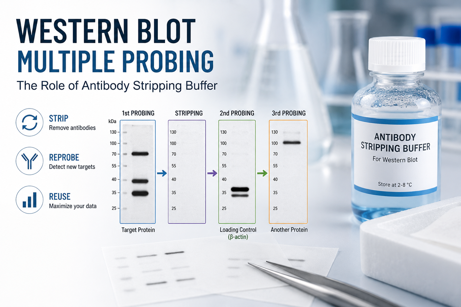
3. Antibody Stripping and Reprobing
Antibody stripping is the process of removing bound primary and secondary antibodies from a Western blot membrane after detection. The membrane is then blocked again and incubated with a new primary antibody. This allows sequential detection of different proteins on the same membrane.
Antibody stripping is useful when membrane cutting is not practical or when multiple targets need to be detected from the same molecular weight region. It is also helpful when sample amount is limited and repeating the Western blot is not ideal.
However, stripping should be used with caution. Each stripping cycle may remove some immobilized protein from the membrane or reduce antigen recognition. For this reason, most laboratories limit stripping to one to three rounds, depending on the membrane type, target abundance, antibody strength, and detection method.
What Is Antibody Stripping Buffer?
Antibody stripping buffer, also known as Western blot stripping buffer, is a reagent designed to disrupt antigen-antibody interactions. Its purpose is to remove previously bound antibodies while keeping the transferred proteins attached to the membrane.
An effective stripping buffer should remove antibody signal completely without causing excessive protein loss. In practice, the balance between stripping efficiency and protein preservation depends on the buffer formulation and incubation conditions.
There are two general categories of stripping buffers: mild stripping buffers and harsh stripping buffers.

Mild Stripping Buffer
Mild stripping buffers often use low pH conditions to weaken antibody binding. Common components may include glycine, SDS at low concentration, and detergent such as Tween-20. These buffers are usually applied at room temperature for a relatively short incubation time.
Mild stripping is preferred for most routine Western blot reprobing because it is less damaging to membrane-bound proteins. It is suitable when the first signal is not extremely strong and when the next target may be low abundance or sensitive to harsh treatment.
Harsh Stripping Buffer
Harsh stripping buffers typically contain stronger denaturing agents such as SDS and reducing agents such as β-mercaptoethanol or DTT. They may also require incubation at elevated temperature.
Harsh stripping is more effective for removing strong antibody signals, but it also increases the risk of protein loss and reduced signal in later probing rounds. It should generally be used only when mild stripping is insufficient.
Recommended Workflow for Stripping and Reprobing
A typical Western blot stripping and reprobing workflow includes the following steps.
First, complete the initial antibody detection and image acquisition. Save all exposure settings and raw data before stripping, because the first signal cannot be recovered once antibodies are removed.
Next, wash the membrane thoroughly with TBST to remove residual detection reagents. Incubate the membrane with the selected antibody stripping buffer according to the manufacturer’s protocol. Mild stripping may take 10 to 30 minutes at room temperature, while harsh stripping may involve heating.
After stripping, wash the membrane multiple times with TBST. This step is critical because residual stripping buffer can interfere with subsequent antibody binding.
To confirm successful stripping, incubate the membrane with secondary antibody only and perform detection. If no signal appears, the previous antibody complex has been adequately removed. If bands remain, additional stripping may be required, or a stronger stripping condition may be needed.
Once stripping is confirmed, block the membrane again using an appropriate blocking buffer, such as 5% non-fat milk or BSA. Then proceed with the next primary antibody incubation, secondary antibody incubation, washing, and detection.
Choosing the Correct Detection Order
Detection order is one of the most important factors in successful multiple probing. In general, low-abundance proteins should be detected before high-abundance proteins. Phosphorylated proteins should also be detected before total proteins or loading controls whenever possible.
A recommended order is:
Phosphorylated protein
Low-abundance target protein
Total target protein
High-abundance loading control
Loading controls such as GAPDH, β-actin, and tubulin are often highly abundant and produce strong signals. If they are detected first, residual signal may remain even after stripping, making later detection more difficult. Strong signals can also increase background and complicate quantitative analysis.
PVDF vs. Nitrocellulose Membranes
The type of membrane also affects stripping performance. PVDF membranes are generally more durable and better suited for repeated stripping and reprobing. They have strong protein-binding capacity and tolerate harsher conditions better than nitrocellulose membranes.
Nitrocellulose membranes can also be stripped, but they are more fragile and may lose protein more easily during repeated washes or harsh stripping. If multiple rounds of reprobing are planned, PVDF is often the preferred membrane.
Common Problems and Troubleshooting
Incomplete stripping is one of the most common issues. If bands from the previous detection remain, extend the stripping time slightly, repeat the stripping step, or use a stronger buffer. Always confirm by secondary-only detection before moving to the next primary antibody.
Weak signal after reprobing may result from protein loss, damaged epitopes, insufficient blocking, or antibody incompatibility. To minimize this problem, use mild stripping conditions whenever possible and avoid unnecessary stripping cycles.
High background after stripping may occur if the membrane is not washed thoroughly or if blocking is insufficient. Additional TBST washes and fresh blocking buffer can help reduce background.
Unexpected bands may be caused by residual antibodies, cross-reactivity, or overlapping molecular weights. Proper controls and careful selection of antibody host species can reduce misinterpretation.
Best Practices for Reliable Multiple Probing
For the best results, plan the entire Western blot strategy before starting the experiment. Choose membrane cutting or multiplex detection whenever possible, because these approaches reduce the need for stripping. Use antibody stripping only when sequential detection on the same membrane is necessary.
Always detect weak or sensitive targets first. Use mild stripping buffer before trying harsh stripping conditions. Validate stripping efficiency before reprobing. Limit the number of stripping cycles, especially when performing quantitative analysis. When possible, keep exposure times within the linear detection range to avoid saturated bands.
It is also important to record all stripping conditions, including buffer type, incubation time, temperature, membrane type, antibody dilution, and detection method. These details make troubleshooting easier and improve experimental reproducibility.
Conclusion
Multiple probing in Western blot is a practical and efficient way to detect several proteins from the same membrane. Researchers can use membrane cutting, multiplex detection, or antibody stripping depending on the molecular weights of the targets, antibody compatibility, and experimental goals.
Antibody stripping buffer provides a useful solution when the same membrane must be reprobed sequentially. Mild stripping buffers are suitable for most applications, while harsh stripping buffers may be used for strong or persistent antibody signals. To obtain reliable results, researchers should optimize stripping conditions, confirm complete antibody removal, and detect low-abundance or phosphorylation-specific targets before high-abundance loading controls.
When performed carefully, Western blot stripping and reprobing can save samples, reduce experimental variation, and improve the efficiency of protein expression analysis.
FireGene, light your research with passion, innovation, and profession.


