Introduction
Kidney research is entering a new era driven by single-cell technologies, spatial biology, and multi-omics analysis. Over the past few years, Single-Cell RNA Sequencing (scRNA-seq), Spatial Transcriptomics, kidney organoids, and AI-assisted bioinformatics have fundamentally transformed how scientists investigate kidney development, injury, regeneration, and disease progression.
Researchers studying Chronic Kidney Disease (CKD), Diabetic Kidney Disease (DKD), Acute Kidney Injury (AKI), Lupus Nephritis, IgA Nephropathy, and Renal Fibrosis are increasingly relying on single-cell approaches to identify disease-driving cell populations, characterize immune responses, and uncover novel therapeutic targets.
However, despite the sophistication of modern sequencing platforms, one reality remains unchanged:
The quality of downstream data is only as good as the quality of the starting cell suspension.
Poor tissue dissociation can result in:
- Low cell viability
- Cell aggregation
- Loss of rare cell populations
- Increased ambient RNA contamination
- Biased sequencing results
- Reduced reproducibility
For this reason, kidney tissue dissociation has become one of the most important upstream steps in modern nephrology research.
This article explores current challenges, workflows, and best practices for preparing kidney tissue for single-cell analysis while highlighting the growing importance of standardized dissociation solutions.
Why Kidney Tissue Is Challenging to Process
Compared with many other tissues, the kidney presents unique difficulties for single-cell isolation.
Complex Cellular Diversity
The kidney contains numerous highly specialized cell populations, including:
- Podocytes
- Mesangial cells
- Proximal tubular epithelial cells
- Distal tubular epithelial cells
- Collecting duct cells
- Endothelial cells
- Fibroblasts
- Resident immune cells
- Infiltrating inflammatory cells
Successful dissociation must preserve representation across all major populations.
Dense Extracellular Matrix
Renal tissue contains substantial extracellular matrix components that become even more abundant during fibrosis.
This makes efficient cell release more difficult than in many soft tissues.
High Cellular Sensitivity
Many kidney cell types are extremely sensitive to:
- Prolonged digestion
- Mechanical stress
- Temperature fluctuations
- Processing delays
Even minor deviations can reduce viability and alter gene expression profiles.
Why Single-Cell Kidney Research Is Growing Rapidly
The rapid adoption of single-cell technologies is being driven by several major research areas.
Diabetic Kidney Disease (DKD)
DKD remains one of the leading causes of kidney failure worldwide.
Recent single-cell studies have identified previously unknown tubular epithelial cell states, immune interactions, and fibrotic pathways that contribute to disease progression.
Acute Kidney Injury (AKI)
Single-cell sequencing allows researchers to examine injury and repair processes at unprecedented resolution.
Researchers can now track:
- Injured epithelial cells
- Regenerative populations
- Inflammatory infiltrates
- Fibrotic transitions
throughout disease progression.
Renal Fibrosis
Fibrosis is considered a common endpoint for many kidney diseases.
Modern single-cell approaches have revealed diverse fibroblast populations with distinct biological functions and therapeutic relevance.
Kidney Organoids
Kidney organoids are increasingly used for:
- Drug discovery
- Disease modeling
- Precision medicine
- Regenerative medicine
These systems require reliable dissociation methods for downstream characterization and sequencing.
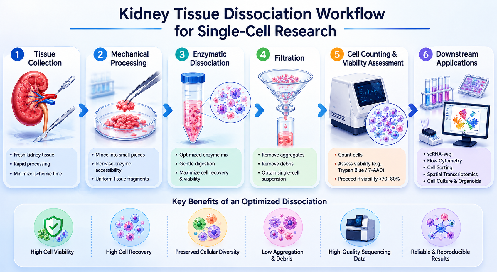
Example Kidney Dissociation Protocol for Single-Cell Research
While exact conditions vary by tissue type and experimental goals, most workflows follow a similar structure.
Step 1 – Tissue Collection
Fresh tissue should be collected promptly and processed rapidly to preserve cellular integrity.
Key Objective
Minimize ischemic injury and RNA degradation.
Step 2 – Mechanical Processing
Kidney tissue is typically minced into small fragments to increase enzyme accessibility.
Key Objective
Create uniform tissue pieces while minimizing mechanical damage.
Step 3 – Enzymatic Dissociation
Specialized enzyme formulations are used to release viable single cells from the tissue matrix.
Key Objective
Maximize cell recovery while preserving sensitive populations.
Step 4 – Filtration
Cell suspensions are filtered to remove:
- Aggregates
- Debris
- Undigested tissue fragments
Key Objective
Generate a clean single-cell suspension.
Step 5 – Cell Counting and Viability Assessment
Before sequencing, researchers typically evaluate:
- Cell concentration
- Viability
- Aggregate content
For many scRNA-seq workflows, cell viability above 70–80% is generally preferred.
Step 6 – Downstream Applications
Prepared cells may be used for:
- Single-Cell RNA Sequencing
- Flow Cytometry
- Cell Sorting
- Cell Culture
- Spatial Biology Workflows
- Multi-Omics Studies
Common Challenges in Kidney Tissue Dissociation
Many sequencing failures can be traced back to sample preparation.
Low Cell Viability
Caused by:
- Excessive digestion
- Delayed processing
- Improper handling
Consequences include poor sequencing performance and reduced data quality.
Cell Aggregation
Cell clumping can reduce effective cell recovery and interfere with microfluidic platforms.
Selective Cell Loss
Certain populations may be more sensitive than others.
This can result in:
- Underrepresentation of rare populations
- Distorted biological conclusions
- Reduced reproducibility
Ambient RNA Contamination
Dead and damaged cells release RNA into the suspension.
This increases background noise and complicates data interpretation.
Case Study: Improving Cell Recovery in a Renal Fibrosis Project
A research group investigating fibrosis progression in a murine CKD model initially experienced poor sequencing performance despite using a leading scRNA-seq platform.
The team observed:
- Low viability
- High debris content
- Reduced recovery of tubular epithelial cells
After reviewing the tissue preparation workflow, the researchers identified dissociation as the primary bottleneck.
By implementing a more standardized tissue processing approach, they achieved:
- Improved cell recovery
- Better viability
- More representative cellular composition
The sequencing platform had never been the problem.
The issue originated during tissue preparation.
This example highlights a common reality in single-cell research:
Data quality begins long before sequencing starts.
Manual Enzyme Cocktails vs Standardized Dissociation Kits
Many laboratories still rely on manually prepared enzyme mixtures.
While these approaches can work, they often introduce variability.
| Factor | Manual Enzyme Cocktail | Standardized Dissociation Kit |
|---|---|---|
| Reproducibility | Variable | Consistent |
| Workflow Complexity | High | Simplified |
| Batch-to-Batch Variation | Higher | Lower |
| Training Requirements | Greater | Reduced |
| Scalability | Limited | Improved |
| Multi-User Consistency | Challenging | Easier |
As projects become larger and more collaborative, standardization becomes increasingly important.
Why Researchers Are Moving Toward Standardized Kidney Dissociation Solutions
Large-scale initiatives such as:
- Kidney Cell Atlas projects
- Multi-center nephrology studies
- Drug discovery programs
- Precision medicine research
require reproducible sample preparation workflows.
Many laboratories therefore prefer kidney-specific solutions designed to support efficient tissue processing.
The FireGene Kidney Dissociation Kit is designed to generate viable single-cell suspensions from kidney tissue for downstream research applications including flow cytometry, cell culture, cell sorting, and single-cell sequencing:
https://firegene.com/products/kidney-dissociation-kit-fg-ba3322?_pos=1&_sid=878daca98&_ss=r
Applications in Spatial Transcriptomics
Spatial transcriptomics is one of the fastest-growing areas in kidney research.
Unlike traditional scRNA-seq, spatial technologies preserve tissue architecture while measuring gene expression.
Many researchers now perform:
- Single-cell RNA sequencing
- Spatial transcriptomics
- Multi-omics validation
using matched samples.
High-quality tissue dissociation remains essential because scRNA-seq datasets are frequently used as references for spatial analyses.
Researchers building integrated workflows often utilize dedicated Kidney Tissue Dissociation Solutions to improve consistency and reproducibility:
https://firegene.com/products/kidney-dissociation-kit-fg-ba3322?_pos=1&_sid=878daca98&_ss=r
Supporting Kidney Organoid Research
Kidney organoids are rapidly becoming indispensable tools for translational research.
Applications include:
- Disease modeling
- Nephrotoxicity testing
- Drug screening
- Personalized medicine
Organoid characterization frequently requires generation of high-quality single-cell suspensions.
Researchers conducting these studies often implement dedicated Single-Cell Kidney Preparation Workflows that support downstream sequencing and phenotypic analysis:
https://firegene.com/products/kidney-dissociation-kit-fg-ba3322?_pos=1&_sid=878daca98&_ss=r
Choosing the Right Kidney Research Supplier
When evaluating a Kidney Research Supplier, investigators should consider:
Tissue-Specific Optimization
Generic solutions may not provide optimal performance for renal tissue.
Workflow Reproducibility
Consistency is increasingly important in multi-center studies.
Compatibility
Support for:
- scRNA-seq
- Flow Cytometry
- Cell Sorting
- Cell Culture
- Organoid Research
- Spatial Transcriptomics
is highly desirable.
Technical Support
Reliable scientific support can significantly reduce troubleshooting time.
For laboratories seeking a Research-Grade Kidney Dissociation Reagent, FireGene provides solutions designed specifically for kidney tissue processing workflows:
https://firegene.com/products/kidney-dissociation-kit-fg-ba3322?_pos=1&_sid=878daca98&_ss=r
Frequently Asked Questions
What is the best method for kidney tissue dissociation?
The optimal method depends on tissue type, disease model, and downstream application. Standardized kidney-specific workflows often improve reproducibility compared with generic enzyme preparations.
How do you prepare kidney tissue for scRNA-seq?
Most workflows involve tissue collection, mechanical disruption, enzymatic dissociation, filtration, viability assessment, and downstream library preparation.
What viability is required for single-cell sequencing?
Many researchers aim for cell viability above 70–80% before sequencing.
Why is kidney tissue difficult to dissociate?
Complex tissue architecture, extracellular matrix content, fibrosis, and cellular sensitivity all contribute to processing challenges.
Can kidney dissociation kits be used for organoid research?
Yes. Many organoid workflows require dissociation for characterization, cell sorting, and sequencing applications.
What causes low cell recovery during kidney tissue processing?
Common causes include excessive digestion, inadequate digestion, mechanical damage, delayed processing, and poor-quality starting tissue.
Future Trends in Kidney Single-Cell Research
Several emerging technologies are expected to further accelerate nephrology research:
- Single-Cell Multi-Omics
- Spatial Proteomics
- AI-Assisted Cell Annotation
- Digital Pathology Integration
- Precision Nephrology Platforms
- Advanced Kidney Organoids
As these technologies mature, sample preparation quality will become even more important.
Researchers increasingly recognize that tissue dissociation is not merely a preparatory step—it is a critical determinant of experimental success.
Conclusion
Single-cell sequencing, spatial transcriptomics, and organoid technologies are revolutionizing kidney research.
From CKD and DKD to AKI, fibrosis, and regenerative medicine, these tools are providing unprecedented insight into renal biology.
Yet every successful dataset begins with one essential requirement:
A high-quality single-cell suspension.
Optimized tissue dissociation improves:
- Cell viability
- Cell recovery
- Reproducibility
- Data quality
For researchers seeking a reliable and standardized solution, the FireGene Kidney Dissociation Kit offers a practical approach for generating viable kidney cell suspensions suitable for modern single-cell and translational research workflows.
References
- Nature Reviews Nephrology. Single-Cell Technologies in Kidney Disease Research.
- Kidney International. Spatial Transcriptomics in Renal Biology.
- Nature Communications. Single-Cell Analysis of Chronic Kidney Disease.
- Journal of the American Society of Nephrology (JASN). Renal Fibrosis and Cellular Heterogeneity.
- Cell Reports Medicine. Kidney Organoids in Precision Medicine.
- Nature Biotechnology. Advances in Single-Cell Sequencing Technologies.
- Nature Reviews Genetics. Multi-Omics Integration in Biomedical Research.
- Clinical Journal of the American Society of Nephrology (CJASN). Emerging Applications of Single-Cell Omics in Nephrology.


