
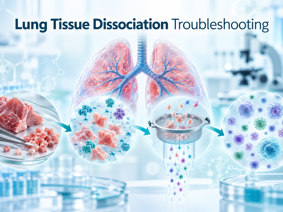
Efficient lung tissue dissociation is a critical first step in many downstream applications, including primary cell culture, flow cytometry, single-cell sequencing, immunophenotyping, organoid establishment, and pulmonary disease research.
However, lung tissue is structurally complex. Alveolar networks, extracellular matrix, blood vessels, mucus, immune cells, and tissue-specific debris can all interfere with enzymatic digestion and single-cell preparation. In pathological lung samples, these challenges may become even more pronounced.
Common problems include incomplete destruction of alveolar structures, low single-cell yield, excessive mucus, red blood cell contamination, poor viability, uneven digestion, and persistent tissue debris.
This article summarizes practical troubleshooting strategies for optimizing lung tissue dissociation and improving the quality of resulting single-cell suspensions.
Why Is Lung Tissue Difficult to Dissociate?
Unlike many relatively homogeneous soft tissues, the lung contains a highly organized three-dimensional architecture.
The tissue includes:
thin alveolar walls;
vascular networks;
connective tissue fibers;
airway structures;
extracellular matrix;
epithelial and endothelial populations;
resident immune cells; and
mucus-producing regions.
Successful dissociation therefore depends not only on enzyme activity but also on tissue preparation, enzyme-to-tissue contact, mechanical disruption, temperature, digestion time, mixing efficiency, and post-digestion processing.
In many cases, simply extending the enzymatic digestion time is not sufficient. If the tissue has not been adequately minced or the alveolar spaces remain physically intact, enzymes may not efficiently reach the relevant structural components.
1. Intact Alveoli Remain After Enzymatic Digestion
The Problem
One of the most common observations during lung dissociation is that a large number of intact alveolar structures remain even after prolonged enzymatic digestion.
The suspension may contain visible tissue fragments or alveolar-like structures, while the final single-cell yield remains very low.
Why It Happens
A major cause is insufficient mechanical mincing before digestion.
If lung tissue fragments remain too large, the alveolar cavities may remain closed. This limits penetration of the dissociation solution and reduces contact between enzymes and the structural components of the alveolar wall.
Increasing digestion time alone may therefore have limited benefit.
How to Improve Alveolar Disruption
A practical optimization strategy includes three steps.
First, mince the lung tissue into very small fragments. For difficult samples, fragments smaller than approximately 0.2 mm³ may improve exposure of internal tissue surfaces.
Second, additional mechanical disruption can be introduced before enzymatic digestion. Repeated puncturing or controlled mechanical disruption may help rupture alveolar structures and increase enzyme accessibility.
Third, if tissue blocks remain visible during digestion, the volume of dissociation solution and digestion duration can be adjusted according to tissue condition.
The central principle is:
Smaller tissue fragments → Better enzyme penetration → More complete structural disruption → Higher single-cell recovery
However, mechanical disruption should remain controlled because excessive shearing may reduce cell viability.
2. How to Handle Viscous Mucus in COPD Lung Samples
The Problem
Lung tissue obtained from chronic obstructive pulmonary disease models may produce a highly viscous suspension during dissociation.
The mucus can trap cells, interfere with pipetting, block filters, and make downstream counting or staining difficult.
Why It Happens
Pathological lung tissue may contain elevated levels of mucus-associated macromolecules. During tissue digestion, these components can mix with cellular debris and extracellular matrix fragments, creating a sticky or gel-like suspension.
Optimization Strategies
One possible approach is to include an appropriate mucopolysaccharide-degrading enzyme when compatible with the downstream application.
Hyaluronidase may be considered in workflows where extracellular glycosaminoglycan-rich material contributes to viscosity, although compatibility with the target cell type should be validated experimentally.
Before filtration, centrifugation can also help separate heavier cellular material from a superficial mucus-rich fraction.
After centrifugation, the upper viscous layer can be carefully removed while retaining the cell-containing fraction.
During filtration, washing the cell strainer with PBS containing serum can help reduce cell retention and improve passage of the suspension through the membrane.
For mucus-rich lung samples, the general workflow becomes:
Digestion → Mucus reduction → Centrifugation → Controlled filtration → Cell recovery
3. Uneven Digestion Caused by Liquid Stratification
The Problem
When lung tissue is digested in a rotating hybridization oven, the suspension may separate into layers.
A relatively clear upper phase and a turbid lower phase may become visible, suggesting that tissue fragments have settled.
Why It Happens
Lung tissue contains vascular material, extracellular matrix, and dense tissue fragments that can sediment when mixing is insufficient.
If tissue remains concentrated at the bottom of the tube, enzyme exposure becomes uneven.
Some fragments may therefore be extensively digested while others remain relatively intact.
How to Improve Mixing
The rotational speed can be moderately increased to improve convection and maintain tissue suspension.
Manual inversion at regular intervals can also improve contact between the dissociation solution and tissue fragments.
In some experimental systems, additional controlled mixing may be introduced, provided that the method does not generate excessive shear stress.
The objective is not vigorous agitation.
Instead, the goal is to maintain:
Uniform tissue suspension + Continuous enzyme contact + Minimal mechanical damage
This balance is particularly important when preparing cells for viability-sensitive downstream applications.
4. How to Determine Whether a Stored Dissociation Solution Has Lost Activity
The Problem
After prolonged storage, a dissociation solution may appear normal but produce poor tissue digestion.
Large tissue blocks remain after incubation, and very few cells are released.
Possible Cause
Repeated freeze-thaw cycles, prolonged storage, or unsuitable storage conditions may reduce enzyme activity.
Because enzyme degradation is not always visually obvious, a functional activity test may be more informative than simply inspecting the solution.
A Small-Scale Digestion Test
A mini digestion experiment can be used before processing valuable samples.
A small quantity of minced lung tissue can be incubated with a defined volume of dissociation solution under standard conditions.
After approximately 30 minutes, gently pipette the sample and observe whether tissue fragments begin to disperse and cells are released.
If tissue pieces remain essentially unchanged and almost no cells are released, the reagent may have lost substantial activity.
For partially active solutions, increasing enzyme volume or digestion time may improve tissue breakdown. However, partially degraded dissociation reagents are generally undesirable for demanding applications such as single-cell RNA sequencing, where cell quality and reproducibility are especially important.
Whenever possible, questionable enzyme preparations should be replaced rather than compensated for with excessively long digestion.
5. Low Viability When Dissociating Neonatal Mouse Lung Tissue
The Problem
Lung tissue from newborn mice may dissociate readily, but the final viability can be substantially lower than expected.
Why Neonatal Lung Is More Sensitive
Immature lung tissue contains relatively fragile cells and less mechanically robust structures than adult tissue.
As a result, neonatal pulmonary cells can be more susceptible to:
prolonged enzymatic exposure;
excessive pipetting;
vigorous shaking;
mechanical shear;
temperature fluctuations; and
insufficient protein protection during washing.
Recommended Adjustments
For neonatal lung tissue, the digestion time should generally be shorter than for adult tissue.
Instead of prolonged digestion, a shorter incubation period followed by gentle evaluation of tissue dissociation can help minimize unnecessary enzyme exposure.
Gentle water-bath incubation may also be preferable to aggressive mechanical agitation.
During washing and resuspension, serum-containing buffer can help reduce mechanical stress and cell loss.
A useful principle is:
Neonatal tissue requires sufficient dissociation, but not maximal dissociation intensity.
The endpoint should be determined by tissue breakup and cell release rather than by applying the longest possible digestion time.
6. Removing Black Particles and Tissue-Derived Debris
The Problem
After centrifugation, dark particles may be visible in or around the cell pellet.
These particles can interfere with automated cell counting, microscopy, flow cytometry, or staining.
Where the Particles Come From
Depending on the tissue source, dark particles may represent:
inhaled environmental material;
carbon-like particulates;
aggregated tissue debris;
blood-derived material; or
insoluble digestion products.
A Simple Separation Strategy
After centrifugation, the pellet can be gently resuspended in a serum-containing buffer.
Allowing the suspension to stand briefly may permit larger or denser particles to settle more rapidly than viable cells.
The upper cell-containing fraction can then be carefully transferred to a fresh tube.
Repeating this procedure can further reduce particulate contamination.
Care should be taken not to extend sedimentation excessively, because desired cells may also begin to settle.
For applications requiring especially clean suspensions, additional density-based or size-based cleanup methods may be evaluated.
7. What to Do After Accidental Over-Digestion
The Problem
If lung tissue is accidentally incubated with dissociation enzymes for several hours, cell viability may fall dramatically.
This is especially common when digestion is performed in a water bath without continuous monitoring.
Why Over-Digestion Is Harmful
Prolonged enzymatic treatment can damage:
cell-surface proteins;
membrane integrity;
extracellular adhesion structures;
receptors used for downstream phenotyping; and
overall cellular viability.
Once substantial damage has occurred, the process cannot be fully reversed.
However, it may still be possible to enrich the remaining viable cell fraction.
Recovery Strategy
Low-speed centrifugation can first be used to remove a portion of light debris and dead-cell material.
The remaining cell fraction can then be washed in serum-containing buffer and centrifuged again under moderate conditions.
After recovery, viability should be reassessed using trypan blue or another suitable viability method.
Cells rescued after severe over-digestion may still be suitable for some culture or exploratory assays, but they may not be appropriate for applications requiring highly intact transcriptomic, surface-marker, or functional profiles.
Prevention therefore remains the preferred strategy.
8. Reducing Endothelial Cell Damage During Red Blood Cell Lysis
The Problem
Lung tissue contains an extensive vascular network, so red blood cell contamination is common after dissociation.
In some samples, erythrocytes may represent a large proportion of the suspension.
Red blood cell lysis can improve purity, but excessive exposure may reduce the viability of sensitive pulmonary vascular endothelial cells.
Why Endothelial Cells May Be Affected
Red blood cell lysis buffers are designed to disrupt erythrocytes selectively, but target-cell tolerance depends on:
buffer formulation;
incubation time;
temperature;
cell type;
sample concentration; and
washing efficiency.
Sensitive primary endothelial cells may be more vulnerable to prolonged or concentrated exposure.
How to Optimize RBC Lysis
When using a red blood cell lysis reagent such as FG-BA3311 Red Blood Cell Lysis Buffer, the lysis step should be kept as short as necessary.
Lower-temperature incubation may help reduce stress on non-erythroid cells.
The reaction should be stopped promptly once red blood cell lysis is sufficient.
Following lysis, cells should be washed thoroughly to remove residual reagent.
If endothelial viability remains poor, a less aggressive lysis condition may be evaluated experimentally by adjusting reagent concentration and incubation time.
For endothelial-focused studies, the aim is not necessarily complete erythrocyte elimination at any cost.
Instead, the goal is:
Adequate RBC removal + Maximum preservation of target-cell viability
Key Variables That Determine Lung Dissociation Quality
Although every tissue sample is different, most lung dissociation problems can be traced to a small number of experimental variables.
Tissue Fragment Size
Smaller fragments improve enzyme penetration but excessive mincing may increase mechanical damage.
Enzyme Exposure
Insufficient digestion leaves tissue clusters, while excessive digestion reduces viability.
Mechanical Force
Controlled mechanical disruption assists dissociation. Excessive pipetting or shaking can damage fragile populations.
Mixing Efficiency
Uniform suspension is essential for consistent enzyme exposure.
Temperature
Temperature affects enzyme activity as well as cellular stress.
Sample Type
Adult, neonatal, fibrotic, COPD, tumor-associated, inflamed, and healthy lung tissues may require different optimization strategies.
Post-Digestion Cleanup
Filtration, RBC removal, washing, debris separation, and centrifugation can strongly influence the final quality of the cell suspension.
A Practical Lung Tissue Dissociation Workflow
A generalized workflow can be summarized as:
Fresh Lung Tissue
↓
Thorough Mechanical Mincing
↓
Controlled Enzymatic Dissociation
↓
Periodic Gentle Mixing
↓
Assessment of Remaining Tissue Structures
↓
Mechanical Dispersion if Necessary
↓
Filtration
↓
Red Blood Cell Removal if Required
↓
Washing and Debris Removal
↓
Cell Counting and Viability Assessment
↓
Downstream Application
At each stage, the procedure should be adapted to the biological properties of the sample rather than applying identical conditions to every specimen.
Applications of High-Quality Lung Single-Cell Suspensions
Optimized lung tissue dissociation can support a wide range of research applications.
These include:
primary lung cell culture;
pulmonary endothelial cell isolation;
epithelial cell studies;
immune-cell profiling;
flow cytometry;
FACS;
single-cell RNA sequencing;
organoid establishment;
pulmonary fibrosis research;
COPD model research;
lung inflammation studies; and
cell-based molecular analysis.
For these applications, the ideal dissociation protocol should provide not only a high cell number but also good viability and preservation of biologically relevant cell populations.
Conclusion
Lung tissue dissociation is a balance between effective structural disruption and preservation of cellular integrity.
Incomplete alveolar breakdown is often related to insufficient tissue mincing and inadequate enzyme penetration. Mucus-rich pathological samples may require additional cleanup strategies, while neonatal tissue generally benefits from gentler and shorter digestion conditions.
Uneven mixing, degraded enzymes, excessive digestion, particulate contamination, and aggressive red blood cell lysis can all reduce the quality of the final cell suspension.
For reliable lung single-cell preparation, researchers should optimize four core factors:
Tissue Preparation + Enzymatic Digestion + Mechanical Handling + Post-Dissociation Cleanup
A troubleshooting-based approach can often improve both single-cell recovery and viability more effectively than simply increasing digestion intensity.
When working with particularly sensitive populations or specialized downstream applications, optimization should be performed using small-scale pilot experiments before processing valuable samples.


