Western blotting (WB) remains one of the most widely used techniques in molecular biology for detecting and quantifying specific proteins. Despite its widespread use, many researchers—especially those new to the technique—encounter persistent issues with high background signals, nonspecific bands, and inconsistent results. A critical but often overlooked step in the Western blot workflow is blocking. Incomplete or improper blocking can severely compromise the quality and interpretability of your data. This blog dives deep into the principles, common problems, and optimization strategies surrounding blocking in Western blot experiments.
What Is Blocking in Western Blotting?
After transferring proteins from an SDS-PAGE gel to a nitrocellulose (NC) or polyvinylidene fluoride (PVDF) membrane, the membrane surface contains exposed sites capable of binding proteins nonspecifically. If left untreated, these sites can attract antibodies during subsequent incubation, leading to high background signals and false-positive bands.
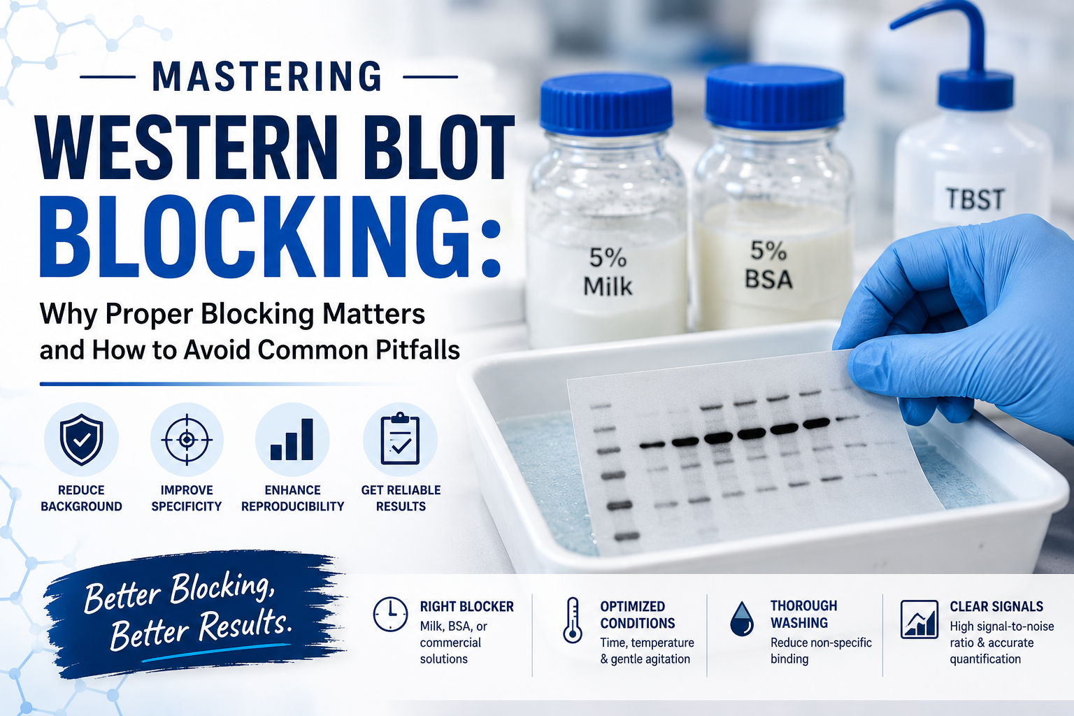
Blocking fills these unoccupied binding sites with an inert protein solution, such as bovine serum albumin (BSA), non-fat dry milk, or commercial blockers. The aim is to minimize nonspecific antibody binding while preserving the accessibility of the target protein for detection.
Common Blocking Agents
1. Non-fat Dry Milk (NFDM)
1. Advantages: Cost-effective, readily available, generally effective for most antibodies.
2. Considerations: Contains casein, which may interfere with detection of phosphorylated proteins or other post-translational modifications.
2. Bovine Serum Albumin (BSA)
1. Advantages: Preferred for detecting phosphorylated or glycosylated proteins, lower background for sensitive applications.
2. Considerations: More expensive than milk, may require optimization for some antibodies.
3. Commercial Blocking Solutions
1. Advantages: Formulated for low background and enhanced reproducibility.
2. Considerations: Higher cost, sometimes requires compatibility testing with specific antibodies.
Consequences of Incomplete Blocking
Failing to properly block the membrane can have several negative effects:

1. High Background Signal
Unblocked sites on the membrane can bind both primary and secondary antibodies nonspecifically. During detection (chemiluminescence, fluorescence, or colorimetric), these nonspecific bindings generate a uniform gray or black background, obscuring the true signal.
2. Nonspecific Bands
Antibodies may attach to proteins other than the target. These bands can appear as extra or unexpected signals and are particularly problematic in complex samples like tissue lysates or serum.
3. Reduced Signal-to-Noise Ratio
High background or extra bands lower the contrast between the target protein and surrounding areas. Weakly expressed proteins may become undetectable or appear diminished.
4. Inaccurate Quantification
Image analysis software may misinterpret background intensity as protein signal, resulting in skewed measurements of protein expression or post-translational modification levels.
5. Poor Reproducibility
Uneven or incomplete blocking leads to variable results across replicates, making it difficult to compare experiments or publish reliable data.
Factors Leading to Incomplete Blocking
Understanding why blocking fails is key to preventing it:
· Insufficient Blocking Time: Short incubation periods may leave many sites unoccupied. Standard protocols recommend 1 hour at room temperature or overnight at 4°C.
· Suboptimal Blocking Concentration: Using a low percentage of BSA or milk may be inadequate for filling all available binding sites.
· Incompatible Blocking Agents: Some antibodies or detection systems are sensitive to components in the blocker. For example, milk contains phosphoproteins that can compete with phospho-specific antibodies.
· Poor Membrane Coverage: Inadequate shaking or insufficient volume during blocking can leave parts of the membrane exposed.
· High Antibody Concentration: Even well-blocked membranes can display nonspecific binding if primary or secondary antibodies are too concentrated.
Optimizing Your Blocking Strategy

1. Choose the Right Blocker
For general applications: 5% non-fat dry milk in TBS-T (Tris-buffered saline with Tween-20) is standard.
For phosphoproteins or sensitive detection: 3–5% BSA in TBS-T is preferred.
2. Adjust Incubation Time and Temperature
Standard room temperature blocking: 1 hour with gentle agitation.
Overnight blocking at 4°C can enhance coverage for challenging membranes or proteins.
3. Ensure Complete Membrane Immersion
Use sufficient volume of blocking solution to cover the entire membrane.
Gentle rocking or orbital shaking ensures uniform exposure.
4. Optimize Antibody Dilution
Start with manufacturer-recommended dilutions and adjust to minimize nonspecific binding.
Too high antibody concentration can overwhelm the blocking layer.
5. Wash Thoroughly After Blocking
Wash with TBS-T to remove unbound blocker before adding primary antibody.
Multiple washes reduce background and improve signal clarity.
6. Consider Sequential Blocking
For especially difficult antibodies or high-background membranes, sequential blocking with different agents (e.g., milk followed by BSA) can sometimes reduce nonspecific binding further.
Practical Tips for Troubleshooting
· Test Multiple Blockers: If background remains high, compare milk, BSA, and commercial blockers side by side.
· Monitor Antibody Specificity: Include a no-primary control to detect secondary antibody–related background.
· Check Membrane Quality: Old or poorly stored PVDF/NC membranes may bind nonspecifically despite proper blocking.
· Document Conditions: Record blocking agent, concentration, duration, and antibody dilutions for reproducibility.
Advanced Considerations
· Fluorescent Western Blotting: Fluorescent detection often demands low autofluorescence blocking agents such as BSA or specialized commercial solutions.
· Multiplex Western Blots: When detecting multiple proteins simultaneously, ensure that the blocker is compatible with all antibodies to prevent cross-reactivity.
· High-Sensitivity Detection: Chemiluminescent or near-infrared detection may exaggerate background if blocking is incomplete; optimize accordingly.
Conclusion
Proper blocking is more than a routine step in Western blotting—it is a critical determinant of data quality. Incomplete blocking leads to high background, nonspecific bands, and poor reproducibility, which can compromise both qualitative and quantitative analysis. By carefully selecting the blocking agent, optimizing incubation conditions, and maintaining thorough washing practices, researchers can significantly improve the clarity and reliability of their Western blot results.
Investing time in blocking optimization ensures that your target protein signal stands out clearly, minimizes false positives, and increases confidence in your experimental conclusions. For every molecular biologist striving for precision, mastering the art of blocking is not optional—it’s essential.
FireGene, light your research with passion, innovation, and profession.


