
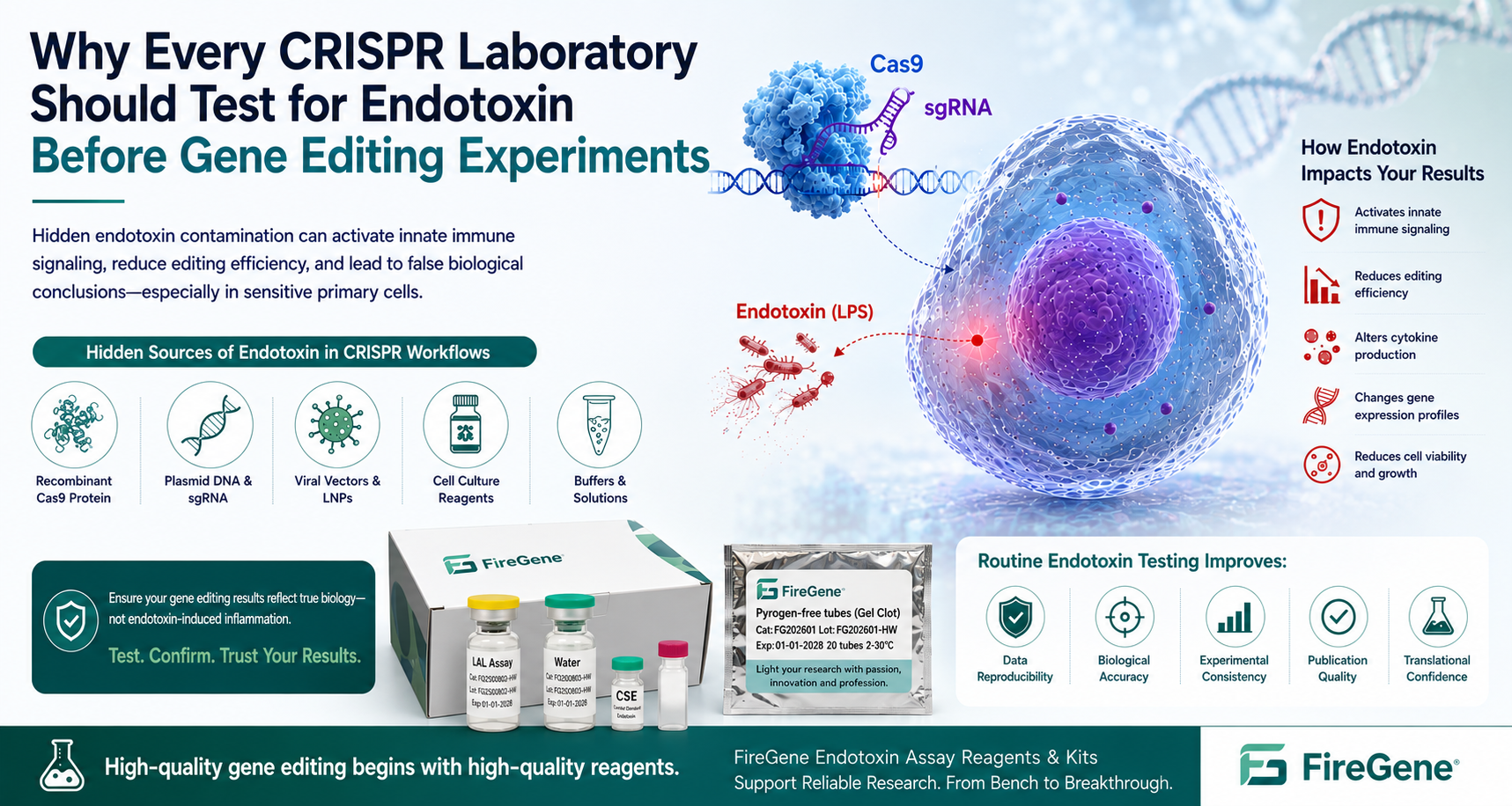
How Hidden Endotoxin Contamination Can Reduce Editing Efficiency, Alter Cellular Responses, and Compromise Experimental Reproducibility
Gene editing technologies have rapidly transformed biomedical research over the past decade. From basic functional genomics to CAR-T cell engineering, stem cell biology, disease modeling, and in vivo therapeutic development, CRISPR-Cas systems have become indispensable tools for laboratories worldwide.
The pace of innovation continues to accelerate.
Today, researchers are no longer limited to traditional CRISPR-Cas9 knockout experiments. Modern gene-editing platforms now include:
- CRISPR-Cas9 genome editing
- CRISPR interference (CRISPRi)
- CRISPR activation (CRISPRa)
- Base editing
- Prime editing
- Epigenome editing
- RNA editing
- Multiplex CRISPR screening
These technologies are being applied across nearly every field of life science, including immunology, oncology, neuroscience, regenerative medicine, agricultural biotechnology, and gene therapy.
However, while laboratories devote enormous effort to optimizing guide RNA design, transfection efficiency, editing specificity, and off-target analysis, one critical experimental variable is still routinely overlooked:
Endotoxin contamination.
Unlike sequencing artifacts or off-target mutations, endotoxin contamination often produces subtle biological effects that are easily mistaken for genuine experimental outcomes.
A CRISPR experiment may appear technically successful:
- Cas9 protein enters the cell.
- Guide RNA is delivered efficiently.
- Editing is confirmed by sequencing.
Yet the downstream phenotype may already have been altered—not by the intended genetic modification, but by bacterial endotoxin introduced through experimental reagents.
This issue is becoming increasingly important because modern gene-editing experiments frequently involve highly sensitive biological systems, including:
- Primary human immune cells
- Peripheral blood mononuclear cells (PBMCs)
- Hematopoietic stem cells
- Induced pluripotent stem cells (iPSCs)
- Organoids
- Mesenchymal stem cells (MSCs)
- CAR-T manufacturing workflows
These cell types are exceptionally responsive to inflammatory stimuli.
Even trace amounts of endotoxin can activate innate immune signaling, alter cytokine secretion, influence cell survival, and modify transcriptional programs—changes that may be incorrectly attributed to CRISPR-mediated gene editing.
For researchers seeking reproducible and biologically meaningful results, endotoxin testing should not be viewed as a quality-control procedure reserved for pharmaceutical manufacturing.
It should be considered an essential component of every well-designed CRISPR experiment.
CRISPR Research Is Entering a New Era
The rapid expansion of CRISPR technologies has fundamentally changed how researchers investigate gene function.
Only a decade ago, many laboratories focused primarily on generating gene knockouts in immortalized cell lines.
Today, CRISPR workflows have become dramatically more sophisticated.
Current research applications include:
- Functional genomic screening
- Precision oncology
- Cell therapy development
- Gene correction for inherited diseases
- Immune engineering
- Synthetic biology
- Developmental biology
- Organoid engineering
- Spatial functional genomics
At the same time, the biological complexity of experimental systems has increased substantially.
Rather than using highly robust transformed cell lines, researchers increasingly perform gene editing in delicate primary cells that more accurately represent physiological biology.
Unfortunately, these same cells are also considerably more sensitive to endotoxin exposure.
Consequently, experimental quality is no longer determined solely by editing efficiency.
Researchers must also consider whether the observed biological response reflects:
- successful genome editing,
or
- unintended activation of inflammatory pathways.
Distinguishing between these two possibilities has become one of the most important quality challenges in modern gene-editing research.
The Hidden Variable That Many CRISPR Laboratories Never Measure
When troubleshooting unsuccessful gene-editing experiments, researchers commonly investigate:
- sgRNA design
- Cas9 activity
- PAM accessibility
- Transfection efficiency
- Cell viability
- DNA repair pathways
- Off-target editing
Rarely does the troubleshooting process include a simple question:
Were the reagents endotoxin-free?
This omission is understandable.
Unlike microbial contamination, endotoxin contamination is invisible.
A recombinant Cas9 preparation may appear perfectly clear.
A plasmid DNA preparation may have acceptable purity ratios.
A guide RNA solution may perform well during quality-control analysis.
Yet all three reagents may still contain biologically significant amounts of endotoxin.
Because endotoxin does not affect reagent appearance, concentration, or sequencing quality directly, contamination frequently goes unnoticed until unexpected biological responses begin appearing in downstream experiments.
These responses often include:
- Increased cytokine production
- Reduced editing efficiency
- Lower cell viability
- Activation of stress-response genes
- Unexpected transcriptional signatures
- Altered differentiation pathways
Without endotoxin testing, researchers may spend weeks optimizing CRISPR protocols while the true source of variability remains undetected.
Why CRISPR Experiments Are Particularly Vulnerable to Endotoxin
Gene-editing workflows typically involve multiple biological reagents that may introduce endotoxin.
Common examples include:
- Recombinant Cas9 protein
- Plasmid DNA
- In vitro-transcribed guide RNA
- Viral vectors
- Lipid nanoparticles
- Electroporation buffers
- Cell culture supplements
- Cytokines
- Growth factors
Each component represents a potential source of lipopolysaccharide (LPS) contamination derived from Gram-negative bacteria.
Even if individual reagents contain only trace amounts of endotoxin, cumulative exposure throughout the workflow may be sufficient to activate cellular immune responses.
This risk becomes especially important when working with highly responsive primary cells.
For example, monocytes and macrophages express Toll-like receptor 4 (TLR4), allowing them to recognize endotoxin rapidly and initiate inflammatory signaling cascades.
Similarly, dendritic cells, PBMCs, and certain stem-cell populations may exhibit profound transcriptional changes following minimal endotoxin exposure.
In these systems, observed phenotypes may no longer reflect genome editing alone—they may instead represent the combined effects of genetic modification and endotoxin-induced immune activation.
Why This Matters More Than Ever
The growing adoption of CRISPR-based therapeutics, cell therapies, and precision medicine has raised expectations for experimental reproducibility.
Academic laboratories, biotechnology companies, and pharmaceutical manufacturers increasingly rely on CRISPR data to support:
- Target validation
- Drug discovery
- Biomarker identification
- IND-enabling studies
- Translational research
- Clinical manufacturing
As these applications move closer to the clinic, unnoticed endotoxin contamination becomes more than a laboratory nuisance.
It becomes a potential source of misleading scientific conclusions.
Increasingly, leading research organizations recognize that validating the biological quality of CRISPR reagents should include not only assessments of purity, concentration, and editing activity, but also routine endotoxin testing using validated bacterial endotoxin assays.
By incorporating endotoxin testing early in the experimental workflow, laboratories can reduce variability, improve reproducibility, and increase confidence that observed phenotypes truly result from gene editing rather than unintended inflammatory activation.
Hidden Sources of Endotoxin in CRISPR Gene Editing Workflows
One of the greatest challenges in endotoxin control is that contamination rarely originates from a single source.
Instead, endotoxin may be introduced gradually throughout the gene-editing workflow, often from reagents that are not routinely tested during academic research.
Unlike GMP manufacturing environments, many research laboratories assume that molecular biology-grade reagents are suitable for all downstream applications. While these reagents may be free from viable microorganisms, they are not necessarily endotoxin-free.
Understanding where endotoxin enters a CRISPR workflow is the first step toward preventing misleading experimental results.
Recombinant Cas9 Protein
Purified Cas9 protein is one of the most widely used components in modern gene editing.
Most commercial Cas9 proteins are produced using recombinant bacterial expression systems, particularly Escherichia coli.
Because endotoxin is a lipopolysaccharide (LPS) found in the outer membrane of Gram-negative bacteria, recombinant protein purification presents an inherent contamination risk.
Even highly purified proteins may retain trace amounts of endotoxin if appropriate removal and quality-control procedures are not implemented.
For many immortalized cell lines, these trace levels may produce little observable effect.
However, for sensitive primary cells, the consequences can be substantial.
Potential effects include:
- Reduced cell viability
- Activation of inflammatory signaling
- Altered cytokine secretion
- Increased experimental variability
- Changes in transcriptional profiles independent of genome editing
These responses may easily be misinterpreted as consequences of successful CRISPR editing rather than reagent contamination.
Plasmid DNA Preparation
Plasmid DNA remains a cornerstone of many CRISPR workflows.
Researchers routinely use plasmids for:
- Cas9 expression
- sgRNA expression
- Donor templates
- Reporter constructs
- Base editors
- Prime editors
Because plasmids are almost universally amplified in E. coli, endotoxin contamination becomes one of the most common quality concerns during plasmid preparation.
Standard molecular biology purification methods often produce DNA of sufficient purity for PCR and cloning.
However, these methods may not adequately remove endotoxin for applications involving:
- Primary immune cells
- Stem cells
- In vivo delivery
- Therapeutic development
Many commercial endotoxin-free plasmid preparation kits have therefore become standard tools in laboratories performing sensitive CRISPR experiments.
Nevertheless, verification through endotoxin testing remains the most reliable method for confirming reagent quality.
Guide RNA (sgRNA)
Synthetic guide RNAs generally present lower endotoxin risk than bacterially produced reagents.
However, contamination may still occur during:
- Manufacturing
- Buffer preparation
- Sample handling
- Laboratory storage
- Reconstitution procedures
In vitro-transcribed guide RNAs may present additional challenges depending on purification methodology.
Although guide RNA itself does not contain endotoxin, improper handling or contaminated reagents may introduce LPS into the final preparation.
Viral Vector Production
Viral vectors continue to play a central role in CRISPR delivery.
Common systems include:
- Lentivirus
- Adeno-associated virus (AAV)
- Adenovirus
Production workflows involve numerous biological reagents, including plasmids, producer cell lines, transfection reagents, purification buffers, and cell culture media.
Each manufacturing stage introduces another opportunity for endotoxin contamination.
Because viral vectors are frequently used for:
- Stem-cell engineering
- Animal studies
- Preclinical development
- Gene therapy research
careful endotoxin monitoring becomes increasingly important throughout production.
Lipid Nanoparticle (LNP) Formulations
Lipid nanoparticles have emerged as one of the fastest-growing delivery technologies for both CRISPR and mRNA therapeutics.
Following the success of mRNA vaccines, LNP-mediated delivery is now widely used for:
- Cas9 mRNA
- Guide RNA
- Base editors
- Prime editors
- Gene silencing
Although lipid formulations themselves are chemically synthesized, contamination may arise from:
- Raw materials
- Buffer components
- Water systems
- Manufacturing equipment
- Laboratory handling
Because LNPs efficiently deliver their cargo into cells, endotoxin contamination within the formulation may likewise be delivered directly into highly sensitive target cells.
Cell Culture Reagents
Many researchers focus exclusively on gene-editing reagents while overlooking the culture environment itself.
Potential endotoxin sources include:
- Fetal bovine serum (FBS)
- Cytokines
- Growth factors
- Albumin preparations
- Cell culture supplements
- Extracellular matrix proteins
Although suppliers implement quality-control procedures, lot-to-lot variability may still occur.
Routine qualification of critical reagents helps minimize unexpected experimental variability.
How Endotoxin Alters CRISPR Experimental Results
One reason endotoxin contamination is particularly problematic is that its biological effects often resemble the phenotypes researchers are attempting to study.
Instead of producing obvious experimental failure, endotoxin frequently generates subtle biological changes that appear scientifically meaningful.
Consequently, contamination may remain undetected throughout an entire project.
Activation of Innate Immune Signaling
The primary cellular receptor for bacterial endotoxin is Toll-like receptor 4 (TLR4).
Upon recognition of LPS, cells rapidly activate downstream inflammatory pathways involving:
- MyD88
- TRIF
- NF-κB
- MAPK
- IRF signaling
These pathways regulate hundreds of genes involved in inflammation and immune activation.
The resulting transcriptional changes may overlap with pathways being investigated in CRISPR studies, making interpretation considerably more difficult.
Increased Cytokine Production
Endotoxin exposure frequently induces production of inflammatory cytokines, including:
- TNF-α
- IL-1β
- IL-6
- IL-8
- MCP-1
- IFN-related mediators
Researchers studying immune regulation may incorrectly attribute these cytokine responses to successful gene editing rather than inadvertent endotoxin exposure.
This represents one of the most common sources of false-positive biological conclusions in immunology research.
Reduced Editing Efficiency
Cellular stress influences virtually every aspect of CRISPR-mediated genome editing.
Endotoxin-induced inflammation may reduce editing performance by:
- Decreasing cell viability
- Altering cell-cycle progression
- Increasing apoptosis
- Reducing proliferation
- Affecting DNA repair pathways
As a result, laboratories may incorrectly conclude that poor editing efficiency reflects problems with guide RNA design or Cas9 activity when the underlying cause is compromised cell health.
Changes in Global Gene Expression
Modern CRISPR studies frequently rely on transcriptomic analysis to characterize edited cells.
However, endotoxin itself induces widespread transcriptional remodeling.
Genes associated with:
- Inflammation
- Stress responses
- Cell survival
- Metabolism
- Immune activation
may become differentially expressed independent of any genomic modification.
Without appropriate endotoxin control, distinguishing genuine editing-induced biology from contamination-induced biology becomes increasingly difficult.
Why Primary Cells Are Especially Sensitive
Not all cell types respond equally to endotoxin.
Immortalized cell lines often tolerate low levels of LPS without obvious phenotypic changes.
Primary cells, however, behave very differently.
This is particularly relevant because many cutting-edge CRISPR applications now involve highly sensitive primary cell systems rather than transformed laboratory cell lines.
The greater the physiological relevance of the experimental model, the greater the importance of endotoxin control.
Peripheral Blood Mononuclear Cells (PBMCs)
PBMCs are among the most endotoxin-sensitive cell populations used in biomedical research.
Even very low endotoxin concentrations can rapidly trigger immune activation, cytokine secretion, and widespread transcriptional changes.
When PBMCs are edited using CRISPR technologies, uncontrolled endotoxin contamination may influence:
- T-cell activation
- Monocyte behavior
- Cytokine release
- Cell proliferation
- Functional immune assays
Consequently, endotoxin testing should be considered an essential quality-control step whenever CRISPR experiments involve human PBMCs.
Why Stem Cells, CAR-T Cells, and Organoids Demand Stricter Endotoxin Control
As CRISPR technology moves beyond conventional cell lines into translational and therapeutic research, the consequences of endotoxin contamination become increasingly significant.
Many next-generation gene-editing applications involve highly sensitive cell populations that respond rapidly to even minimal inflammatory stimulation.
Unlike immortalized cell lines such as HEK293 or HeLa cells, these models often possess intact innate immune signaling pathways and maintain physiological responses that closely resemble those observed in vivo.
Consequently, endotoxin contamination may fundamentally alter experimental outcomes before genome editing can even be properly evaluated.
Induced Pluripotent Stem Cells (iPSCs)
Human induced pluripotent stem cells (iPSCs) have become one of the most important CRISPR research models.
Applications include:
- Disease modeling
- Gene correction
- Developmental biology
- Drug screening
- Regenerative medicine
However, iPSCs are notoriously sensitive to environmental stress.
Even modest inflammatory stimulation may influence:
- Colony morphology
- Self-renewal
- Cell survival
- Differentiation potential
- Epigenetic stability
Researchers frequently spend months optimizing differentiation protocols.
If endotoxin contamination occurs during genome editing, the resulting changes in lineage commitment may mistakenly be attributed to successful gene modification rather than activation of inflammatory pathways.
For this reason, many stem-cell facilities now recommend using only endotoxin-controlled reagents throughout CRISPR workflows.
Hematopoietic Stem Cells
Gene editing of hematopoietic stem cells (HSCs) has become central to developing treatments for:
- Sickle cell disease
- β-thalassemia
- Primary immunodeficiencies
- Fanconi anemia
- Various inherited blood disorders
Unlike continuously growing laboratory cell lines, HSCs possess limited proliferative capacity and exhibit considerable sensitivity to culture conditions.
Endotoxin exposure may influence:
- Stem-cell viability
- Engraftment potential
- DNA repair efficiency
- Long-term self-renewal
These variables directly affect both research outcomes and translational development.
CAR-T Cell Engineering
CRISPR is increasingly used during CAR-T manufacturing to:
- Knock out immune checkpoint genes
- Eliminate endogenous TCR expression
- Improve persistence
- Enhance tumor specificity
- Reduce graft-versus-host disease
Throughout this process, T cells undergo multiple activation, expansion, and genetic engineering steps.
Endotoxin contamination during any stage may alter:
- T-cell activation kinetics
- Cytokine production
- Cellular exhaustion
- Expansion efficiency
- Functional cytotoxicity
Because cytokine secretion represents a major endpoint in CAR-T research, distinguishing genuine CRISPR-induced immune responses from endotoxin-induced activation becomes critically important.
Organoid Models
Brain, intestinal, liver, kidney, and lung organoids are increasingly used to investigate human development and disease.
Compared with traditional monolayer cultures, organoids possess complex multicellular architecture and closely resemble native tissue.
However, they also demonstrate heightened sensitivity to inflammatory stress.
Endotoxin contamination may affect:
- Organoid maturation
- Cellular composition
- Morphological development
- Stem-cell niche maintenance
- Gene expression profiles
Researchers studying developmental pathways may therefore generate misleading conclusions if reagent quality is not carefully controlled.
Acceptable Endotoxin Levels for CRISPR Gene Editing Experiments
One of the most frequently asked questions is:
"What endotoxin level is acceptable for CRISPR experiments?"
The answer is less straightforward than many researchers expect.
Unlike pharmaceutical manufacturing, there is no universal endotoxin specification applicable to every research experiment.
The acceptable endotoxin level depends on multiple factors, including:
- Cell type
- Gene-editing strategy
- Exposure duration
- Delivery system
- Downstream assay
- Experimental objective
For example, an endotoxin concentration that produces no measurable response in HEK293 cells may significantly activate human PBMCs.
Similarly, concentrations tolerated during routine plasmid transfection may prove unacceptable for stem-cell engineering or CAR-T manufacturing.
Because of these differences, laboratories should validate endotoxin limits according to their specific biological system rather than relying on a single universal threshold.
Relative Sensitivity of Common CRISPR Models
In general, sensitivity follows approximately this pattern:
Very High Sensitivity
- Human PBMCs
- Monocytes
- Macrophages
- Dendritic cells
- iPSCs
- Hematopoietic stem cells
High Sensitivity
- Primary T cells
- Primary neurons
- Brain organoids
- Liver organoids
- Mesenchymal stem cells
Moderate Sensitivity
- Primary fibroblasts
- Endothelial cells
- Primary epithelial cells
Lower Sensitivity
- HEK293
- HeLa
- CHO cells
- Other immortalized cell lines
Even within the same cell type, donor variability, activation state, and culture conditions may substantially influence endotoxin responsiveness.
Why Validation Matters More Than a Universal Number
Rather than asking,
"What endotoxin level is considered safe?"
a better scientific question is:
"At what endotoxin concentration does my experimental model begin showing measurable biological changes?"
Researchers should evaluate:
- Cell viability
- Cytokine secretion
- Editing efficiency
- Gene expression
- Functional phenotype
under their own experimental conditions.
This validation-based approach aligns with current best practices in both academic research and translational development.
Why Every CRISPR Laboratory Should Implement Routine Endotoxin Testing
Many laboratories routinely verify:
- DNA concentration
- RNA integrity
- Protein purity
- Cell viability
- Mycoplasma status
Yet endotoxin testing is often omitted despite its ability to influence virtually every downstream biological assay.
Routine endotoxin testing offers several important advantages.
It helps laboratories:
- Identify contaminated reagent lots before experiments begin.
- Improve reproducibility between projects.
- Reduce unexplained experimental variability.
- Prevent false-positive inflammatory phenotypes.
- Increase confidence in published results.
- Support future translational development.
Perhaps most importantly, endotoxin testing enables researchers to distinguish between biology caused by genome editing and biology caused by bacterial contaminants.
Choosing the Right Endotoxin Testing Method
Several validated approaches are available for endotoxin detection, each suited to different laboratory needs.
Common methods include:
- Gel Clot TAL/LAL Reagent — a simple qualitative assay widely used for routine screening.
- Kinetic Chromogenic TAL/LAL Reagent — provides quantitative measurements with high sensitivity and broad dynamic range, making it ideal for research laboratories handling multiple reagent types.
- Kinetic Turbidimetric TAL/LAL Reagent — suitable for automated workflows and higher-throughput testing.
For laboratories routinely performing CRISPR experiments involving primary cells, stem cells, organoids, or cell therapy research, incorporating a validated TAL/LAL Reagent workflow before critical experiments can substantially reduce experimental uncertainty.
FireGene provides a comprehensive portfolio of endotoxin testing solutions for research and pharmaceutical laboratories, including Gel Clot and Kinetic Chromogenic TAL/LAL Reagents, Control Standard Endotoxin (CSE), and Endotoxin Assay Water.
Case Study: When Endotoxin—Not CRISPR—Explained the Experimental Phenotype
Consider a research laboratory investigating the role of a novel immune regulatory gene in human macrophages using CRISPR-Cas9.
The experimental workflow appeared technically successful.
Researchers confirmed:
- High Cas9 protein purity
- Efficient sgRNA delivery
- More than 80% gene-editing efficiency
- Excellent sequencing quality
- Expected on-target mutations
However, RNA sequencing revealed an unexpected finding.
Compared with control cells, edited macrophages exhibited dramatic upregulation of inflammatory genes, including:
- TNF
- IL6
- CXCL8
- CCL2
- NFKBIA
Initially, investigators concluded that deletion of the target gene triggered a strong inflammatory phenotype.
The findings appeared biologically plausible and aligned with several published reports.
Before submitting the manuscript, however, the laboratory performed additional quality-control testing on the recombinant Cas9 protein lot.
Unexpectedly, measurable endotoxin contamination was detected.
The researchers repeated the experiment using an endotoxin-controlled Cas9 preparation.
The inflammatory signature largely disappeared.
Editing efficiency remained unchanged.
The only variable that differed between the two experiments was endotoxin contamination.
The apparent immune phenotype had not been caused by CRISPR-mediated gene disruption.
Instead, it resulted from unintended activation of the TLR4–NF-κB signaling pathway by endotoxin introduced during reagent preparation.
Although simplified, this example reflects a challenge increasingly recognized in immunology and gene-editing research.
Without endotoxin testing, contamination may generate convincing—but ultimately misleading—biological conclusions.
A Practical CRISPR Endotoxin Quality Control Checklist
Implementing routine endotoxin control does not require major changes to existing workflows.
Instead, researchers can integrate a few straightforward quality-control practices before initiating gene-editing experiments.
Before Ordering Reagents
✔ Verify whether recombinant proteins are tested for endotoxin.
✔ Request Certificates of Analysis (COAs) whenever available.
✔ Confirm that plasmid DNA preparations are produced using endotoxin-free purification methods.
Before Genome Editing
✔ Test recombinant Cas9 protein when working with sensitive primary cells.
✔ Confirm endotoxin levels in plasmid DNA preparations.
✔ Review endotoxin specifications for viral vectors and delivery reagents.
✔ Use endotoxin-free water and buffers whenever possible.
✔ Avoid repeated freeze-thaw cycles that may introduce contamination through handling.
Before Cell-Based Functional Assays
✔ Confirm high cell viability following editing.
✔ Monitor cytokine release in immune-cell experiments.
✔ Include appropriate negative controls.
✔ Evaluate unexpected inflammatory responses before interpreting biological mechanisms.
✔ Consider routine endotoxin testing as part of standard laboratory quality assurance.
By incorporating these simple practices, laboratories can substantially improve experimental reproducibility while reducing the risk of false biological interpretations.
Why Endotoxin Testing Should Become Standard Practice in CRISPR Laboratories
As CRISPR technology continues expanding into translational medicine, quality expectations are rapidly evolving.
Funding agencies, scientific journals, and biotechnology companies increasingly emphasize:
- Experimental reproducibility
- Transparent reporting
- Robust quality control
- Reliable biological interpretation
Routine endotoxin testing supports all four objectives.
Rather than being viewed solely as a pharmaceutical manufacturing requirement, endotoxin testing is becoming an important research quality-control tool—particularly for laboratories working with:
- Primary human cells
- Stem cells
- CAR-T engineering
- Organoids
- Gene therapy vectors
- Functional genomics
- Precision medicine
The cost of a simple endotoxin assay is negligible compared with the time and resources required to repeat months of CRISPR experiments because of an unrecognized contaminated reagent.
Supporting Reliable Gene Editing Research with FireGene Endotoxin Testing Solutions
Reliable CRISPR experiments begin with reliable reagents.
FireGene offers a comprehensive portfolio of TAL/LAL Reagent solutions designed to support endotoxin detection across research, biotechnology, and pharmaceutical laboratories.
Available products include:
- Gel Clot TAL/LAL Reagent
- Kinetic Chromogenic TAL/LAL Reagent
- Kinetic Turbidimetric TAL/LAL Reagent
- Control Standard Endotoxin (CSE)
- Endotoxin Assay Water
- Endotoxin testing accessories
These products are suitable for evaluating recombinant proteins, plasmid DNA preparations, buffers, biological reagents, and other materials commonly used in CRISPR workflows.
Whether performing basic research or developing advanced gene-editing therapeutics, implementing routine endotoxin testing can improve confidence in experimental outcomes and help ensure that observed biological effects truly result from genome editing—not unintended inflammatory contamination.
Frequently Asked Questions (FAQ)
1. Can endotoxin reduce CRISPR editing efficiency?
Yes. Endotoxin-induced cellular stress may reduce cell viability, alter DNA repair activity, and negatively affect overall editing performance, particularly in primary cells and stem cells.
2. Does recombinant Cas9 protein contain endotoxin?
It can. Because recombinant Cas9 is commonly produced in E. coli, trace endotoxin contamination may remain unless appropriate purification and quality-control procedures are performed.
3. Should plasmid DNA be tested for endotoxin?
Yes. Plasmids are typically amplified in bacterial hosts, making endotoxin contamination a common concern. Endotoxin-free plasmid preparation and verification are recommended for sensitive applications.
4. Which CRISPR experiments are most sensitive to endotoxin?
Experiments involving PBMCs, macrophages, dendritic cells, iPSCs, hematopoietic stem cells, CAR-T cells, and organoids are generally much more sensitive than experiments using immortalized cell lines.
5. Is there a universal acceptable endotoxin limit for CRISPR research?
No. Acceptable endotoxin levels depend on the biological model, reagent dosage, exposure duration, and experimental objectives. Each laboratory should establish application-specific acceptance criteria.
6. Can software identify endotoxin contamination?
No. Bioinformatics tools cannot distinguish gene-expression changes caused by endotoxin from those caused by genome editing. Preventive laboratory quality control remains essential.
7. Should CRISPR laboratories routinely test recombinant proteins for endotoxin?
For experiments involving primary cells, stem cells, immune cells, organoids, or therapeutic development, routine endotoxin testing is strongly recommended.
While recombinant proteins are often supplied with purity specifications, protein purity does not necessarily indicate low endotoxin burden. Measuring endotoxin before introducing recombinant proteins into sensitive biological systems helps reduce experimental variability and improves reproducibility.
8. Can endotoxin affect CRISPR screening experiments?
Absolutely.
Genome-wide CRISPR screens often rely on subtle differences in cell proliferation, survival, differentiation, or transcriptional activity.
Endotoxin contamination may introduce systematic bias by:
- Altering immune signaling pathways
- Changing cell growth rates
- Activating stress-response genes
- Modifying apoptosis pathways
Because these changes may occur across entire cell populations, contamination can distort screen results and complicate hit identification.
9. Is endotoxin testing only important for therapeutic research?
No.
Although endotoxin testing is mandatory for many pharmaceutical products, it also provides substantial value in basic research.
Academic laboratories increasingly recognize that eliminating hidden experimental variables improves:
- Data reproducibility
- Publication quality
- Cross-laboratory consistency
- Confidence in biological interpretation
Routine endotoxin testing is becoming a best practice whenever experiments involve biologically sensitive cells.
10. Which endotoxin testing method is most suitable for research laboratories?
The optimal method depends on laboratory workflow and throughput requirements.
Many research laboratories prefer:
- Gel Clot TAL/LAL Reagent for straightforward qualitative screening.
- Kinetic Chromogenic TAL/LAL Reagent for quantitative analysis of multiple reagents.
- Kinetic Turbidimetric TAL/LAL Reagent for higher-throughput workflows.
When selecting an assay, researchers should consider:
- Required sensitivity
- Sample matrix
- Number of samples
- Available instrumentation
- Regulatory or publication requirements
FireGene offers a complete portfolio of endotoxin testing solutions suitable for both research and pharmaceutical applications.
Conclusion
CRISPR technology has fundamentally changed how scientists investigate biology, engineer cells, and develop next-generation therapeutics.
From functional genomics to regenerative medicine, genome editing is now one of the most powerful tools in modern biomedical research.
Yet the success of a CRISPR experiment depends on far more than guide RNA design or editing efficiency.
Every experiment also depends on the biological quality of the reagents introduced into living cells.
Among the many hidden variables that influence gene-editing outcomes, endotoxin contamination remains one of the most underestimated.
Unlike obvious laboratory contamination, endotoxin rarely causes immediate experimental failure.
Instead, it subtly alters cellular physiology by activating innate immune signaling, changing cytokine production, modifying transcriptional programs, and influencing cell survival.
These changes may easily be mistaken for genuine consequences of genome editing.
As CRISPR research increasingly focuses on sensitive biological systems—including primary immune cells, induced pluripotent stem cells, CAR-T cells, hematopoietic stem cells, and organoids—the importance of rigorous endotoxin control will continue to grow.
Rather than relying solely on downstream troubleshooting or computational interpretation, researchers should adopt a proactive quality-control strategy that includes routine endotoxin assessment of critical reagents.
Testing recombinant proteins, plasmid DNA, viral vectors, and other key materials before initiating experiments can dramatically improve reproducibility while reducing the risk of misleading biological conclusions.
In today's era of precision genome engineering, high-quality gene editing begins with high-quality reagents.
By incorporating validated TAL/LAL Reagent testing into routine laboratory workflows, researchers can improve experimental confidence, strengthen publication quality, and ensure that observed phenotypes truly reflect genome editing—not unintended inflammatory activation caused by endotoxin contamination.
Explore the complete range of FireGene Endotoxin Assay Reagents and Kits for reliable endotoxin detection in CRISPR, cell therapy, recombinant protein, and molecular biology research.
References
- Jinek M, Chylinski K, Fonfara I, Hauer M, Doudna JA, Charpentier E. A programmable dual-RNA-guided DNA endonuclease in adaptive bacterial immunity. Science. 2012;337(6096):816–821.
- Cong L, Ran FA, Cox D, et al. Multiplex Genome Engineering Using CRISPR/Cas Systems. Science. 2013;339(6121):819–823.
- Mali P, Yang L, Esvelt KM, et al. RNA-Guided Human Genome Engineering via Cas9. Science. 2013;339(6121):823–826.
- Anzalone AV, Randolph PB, Davis JR, et al. Search-and-replace genome editing without double-strand breaks or donor DNA. Nature. 2019;576:149–157.
- Komor AC, Kim YB, Packer MS, Zuris JA, Liu DR. Programmable editing of a target base in genomic DNA without double-stranded DNA cleavage. Nature. 2016;533:420–424.
- Hendel A, Fine EJ, Bao G, Porteus MH. Quantifying genome-editing outcomes at endogenous loci using CRISPR/Cas9. Nature Biotechnology. 2015;33:985–989.
- Latz E, Xiao TS, Stutz A. Activation and regulation of the inflammasomes. Nature Reviews Immunology. 2013;13:397–411.
- Rietschel ET, Brade H, Holst O, et al. Bacterial Endotoxin: Molecular Relationships of Structure to Activity and Function. FASEB Journal. 1994;8:217–225.
- USP <85> Bacterial Endotoxins Test. United States Pharmacopeia.
- USP <86> Bacterial Endotoxins Test Using Recombinant Reagents. United States Pharmacopeia.
- ICH Q9(R1). Quality Risk Management. International Council for Harmonisation.
- FDA. Guidance for Industry: Pyrogen and Endotoxins Testing: Questions and Answers.
FireGene Endotoxin Testing
Ready to run your endotoxin assay?
FireGene offers a complete endotoxin testing toolkit — from TAL reagents and CSE standards to pyrogen-free consumables and LAL reagent water. All products are aligned with USP <85>, EP 2.6.14, and JP 4.01.


