
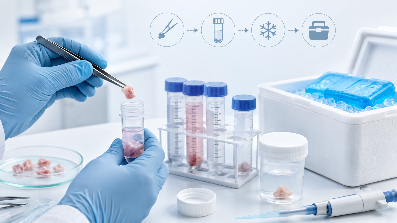
Tissue samples should be collected using clean instruments, handled gently, and preserved immediately through appropriate fixation, freezing, or storage methods based on the intended downstream analysis. A well-collected and properly preserved tissue sample can protect morphology, maintain antigen quality, and support clearer results during downstream testing. This is especially important when the sample will be used for antibody-based detection, where tissue preparation, tissue storage, primary antibody selection, and secondary antibody selection all influence the final signal.
How to collect and preserve tissue samples depends on the purpose of the experiment. A sample intended for DNA analysis may require different handling than one intended for antibody labelling. A tissue section planned for immunohistochemistry may require fixation, embedding, sectioning, antigen retrieval, blocking, and antibody incubation. A sample prepared for frozen sectioning may need rapid freezing and careful tissue storage to protect both morphology and antigen accessibility.
Scientific Basis and Practical Scope
Tissue preparation is a quality-driven process. The way a tissue is collected, fixed, frozen, stored, and sectioned can affect antigen detection and antibody labelling. In immunohistochemistry, tissue sections are commonly processed through antigen retrieval, blocking, primary antibody incubation, secondary antibody detection, and visualisation steps. Antibody-based workflows also require careful antibody validation. Research guidelines emphasise that antibodies should be selected and validated according to the application, sample type, target, and detection method.
The goal is simple: preserve tissue quality, protect antigen integrity, select suitable antibodies, and generate reliable staining with low background and clear target localisation.
Why Tissue Sample Collection and Preservation Matter
Tissue collection and preservation directly affect experimental reliability. If tissue dries, degrades, freezes poorly, or is fixed incorrectly, the final results may be harder to interpret.
Preserving Tissue Morphology
Morphology refers to the structure and appearance of the tissue. Good preservation maintains cellular architecture, which is important for histology, IHC, IF, and cardiac tissue analysis.
Protecting Antigen Integrity
Antigens are the targets recognised by antibodies. Poor preservation can mask, damage, or reduce antigen accessibility. This can lead to weak antibody labelling or inconsistent staining.
Maintaining DNA, RNA, and Protein Quality
Different preservation methods protect different biomolecules. Frozen tissue may support molecular analysis, while fixed tissue may be better for morphology and antibody-based localisation.
Improving Reproducibility
Standardised tissue storage, fixation, sectioning, and antibody selection help reduce variation between samples and experiments.
Types of Tissue Samples
Tissue samples can be collected and preserved in different formats depending on the downstream application.
Fresh Tissue Samples
Fresh tissue is processed shortly after collection. It can be useful for live-cell workflows, enzymatic tissue preparation, or assays where fixation is not desired.
Frozen Tissue Samples
Frozen tissue is useful for molecular analysis, cryosectioning, and some antibody labelling workflows. It requires careful freezing and storage to avoid freeze-thaw damage.
FFPE Tissue Samples
Formalin-fixed paraffin-embedded tissue is widely used in histology and immunohistochemistry. It preserves morphology well, but antigen retrieval may be required because fixation can mask epitopes.
Cardiac Tissue
Cardiac Tissue needs careful handling because muscle architecture, fibrosis, vascular regions, and cell density can affect section quality and staining. Compression, drying, and delayed preservation should be minimised.
Liquid and Body Fluid Samples
Blood, serum, plasma, and body fluids may be preserved for antibody detection, protein analysis, DNA/RNA analysis, or biomarker testing.

General Rules for Tissue Sample Collection
Good tissue collection begins before the sample is removed.
Define the Downstream Assay First
Before collection, decide whether the sample will be used for antibody labelling, DNA/RNA analysis, protein extraction, histology, cardiac tissue analysis, or antibody development process testing. The preservation method should match the final assay.
Use Clean Tools and Containers
Use clean, suitable containers and tools. Avoid contamination, drying, and sample mix-ups. Label each sample clearly.
Avoid Crushing the Tissue
Do not compress tissue with forceps. Crushing can damage morphology and create artefacts that affect tissue preparation and antibody labelling.
Control Time Before Preservation
The time between collection and fixation or freezing should be minimised. Faster preservation helps protect antigen quality and reduces degradation.
Record Sample Metadata
Record tissue source, collection time, preservation method, storage temperature, tissue size, and intended assay. This improves traceability and trust.
Tissue Storage and Transport Conditions
Tissue storage and transport should protect the sample until processing.
Short-Term Refrigerated Storage
For short-term handling, some tissues may be held cold in a suitable medium or container. The storage time and temperature should be validated for the target assay.
Ultra-Low Temperature Storage
Ultra-low-temperature storage may be used for long-term tissue preservation, especially when the sample is intended for molecular testing or frozen tissue workflows.
Dry Ice Transport
Dry ice can support frozen sample transport, but transport containers must be suitable and safe. Samples should be protected from temperature fluctuation and contamination.
Leakproof Containers
Fresh, frozen, or chemically preserved tissues should be transported in secure, leakproof containers. Clear labelling and documentation help prevent delays and sample loss.
Common Tissue Preservation Methods
There is no single best preservation method for every sample. The method should match the tissue type and downstream application.
Cooling and Refrigeration
Cooling is useful for short-term tissue storage when immediate processing is expected. It can slow degradation and preserve sample conditions for a limited time.
Cryogenic and Ultra-Low Temperature Freezing
Freezing is useful for long-term storage and molecular applications. Frozen tissue can be used for cryosections, protein analysis, DNA/RNA work, and some antibody labelling workflows. Repeated freeze-thaw cycles should be avoided.
Chemical Preservation
Chemical preservation includes formalin fixation, alcohol preservation, and other fixative-based approaches. Formalin fixation is common for FFPE tissue preparation. It preserves morphology but may require antigen retrieval for antibody labelling.
Drying and Desiccation
Drying and desiccants may be useful for specific field or DNA-preservation applications. However, dried tissue is usually not ideal for high-quality morphology or antibody labelling.
Liquid Preservation
Blood, serum, plasma, and other liquid samples may require separation, refrigeration, freezing, or stabilising reagents depending on the target analyte.
Tissue Preparation for Antibody Labelling
Antibody labelling depends on both tissue quality and antibody quality.
Fixation
Fixation stabilises tissue structure and helps preserve morphology. However, fixation must be balanced because over-fixation can reduce antigen accessibility.
Embedding and Sectioning
Tissue may be embedded in paraffin or prepared as frozen blocks. Thin, consistent sections help improve staining and imaging.
Antigen Retrieval
Antigen retrieval helps expose epitopes that may be masked by fixation. This step is often important in immunohistochemistry and must be optimised for each antibody and tissue type.
Blocking
Blocking reduces non-specific antibody binding and background signal. It should be matched to the antibody system and tissue type.
Primary Antibody Incubation
The primary antibody binds directly to the target antigen. Its specificity, validation, clone, host species, and concentration all affect staining quality.
Secondary Antibody Detection
The secondary antibody binds to the primary antibody and carries a detection label such as an enzyme or fluorophore. Secondary antibodies are selected based on the host species and isotype of the primary antibody.
Antibody Selection for Tissue Samples
Antibody selection is one of the most important steps in tissue staining.
Choosing the Right Primary Antibody
A primary antibody should be specific to the target, validated for the application, and suitable for the tissue format. For example, an antibody validated for Western blot may not always work well for IHC or IF.
Important checks include:
|
Selection Factor |
Why It Matters |
|
Target specificity |
Confirms the antibody binds the right protein |
|
Application validation |
Supports use in IHC, IF, WB, ELISA, or flow cytometry |
|
Host species |
Determines secondary antibody choice |
|
Clone type |
Affects consistency and binding pattern |
|
Tissue validation |
Confirms staining in relevant sample type |
|
Controls |
Helps confirm true signal vs background |
Choosing the Right Secondary Antibody
The secondary antibody should match the primary antibody host species and isotype. It should also be compatible with the detection system, imaging platform, and tissue species. Cross-adsorbed secondary antibodies can help reduce background in some tissue staining workflows. No-primary controls are also useful for checking secondary antibody background.
Recombinant Antibody Production Methods and Tissue Staining
Recombinant antibody production methods are increasingly important in antibody-based research because they can improve consistency. Recombinant antibodies are produced from defined antibody sequences, which can support better lot-to-lot reproducibility compared with less-defined reagent sources.
Why Recombinant Antibodies Are Useful
Recombinant antibodies can support consistent antibody labelling, long-term reproducibility, and more controlled antibody development process workflows. They can be useful in IHC, IF, ELISA, flow cytometry, and therapeutic antibody development, guiding workflows.
Role in Antibody Development
During the antibody development process, tissue samples may be used to confirm target localisation, evaluate staining pattern, test specificity, and compare signal across tissue types.
Cardiac Tissue Collection and Preservation
Cardiac Tissue requires careful tissue handling because heart muscle has a strong structure and region-specific biology.
Preserving Muscle Architecture
Heart tissue should be handled gently to preserve cardiomyocyte arrangement and tissue morphology.
Avoiding Compression Artefacts
Compression can distort cardiac fibres and affect the interpretation of staining. Use gentle tools and suitable containers.
Freezing Cardiac Tissue
Frozen cardiac tissue may be useful for molecular analysis, protein extraction, or frozen section antibody labelling.
Fixing Cardiac Tissue
Fixed cardiac tissue may be preferred for morphology and IHC. Fixation time, tissue thickness, and antigen retrieval should be optimised for the target marker.
Antibody Labelling Workflow for Preserved Tissue
A practical antibody labelling workflow may include the following steps:
Step 1: Collect and Preserve Tissue
Choose fixation, freezing, chemical preservation, or liquid preservation based on the downstream assay.
Step 2: Embed and Section
Prepare tissue sections using paraffin embedding or frozen sectioning.
Step 3: Prepare the Slide
Deparaffinize FFPE sections or prepare frozen sections according to the protocol.
Step 4: Retrieve Antigen
Use heat-induced or enzyme-based antigen retrieval when needed.
Step 5: Block Non-Specific Binding
Apply blocking buffer to reduce background signal.
Step 6: Apply Primary Antibody
Incubate with a validated primary antibody at an optimised dilution.
Step 7: Apply Secondary Antibody
Use a compatible secondary antibody for detection.
Step 8: Wash, Counterstain, and Image
Wash carefully, counterstain if needed, mount, and image under suitable conditions.
Tissue Storage by Research Application
|
Application |
Best Preservation Focus |
|
DNA analysis |
Preserve nucleic acid quality |
|
RNA analysis |
Rapid stabilization and low-temperature storage |
|
Protein analysis |
Avoid degradation and freeze-thaw cycles |
|
Antibody labeling |
Preserve antigen and morphology |
|
Histology/IHC |
Fixation and antigen retrieval compatibility |
|
Flow cytometry |
Maintain cell quality when live cells are required |
|
Cardiac tissue research |
Protect muscle architecture and target antigens |
|
Antibody development |
Confirm target localization and specificity |
Common Mistakes During Tissue Collection and Preservation
Common mistakes include delayed fixation, tissue drying, incorrect storage method, missing sample metadata, tissue compression, repeated freeze-thaw cycles, and the use of antibodies not validated for the tissue format. These issues can be reduced through clear SOPs, validated preservation methods, proper tissue preparation, and careful antibody selection.
Troubleshooting Tissue Preservation and Antibody Labelling
|
Problem |
Possible Cause |
Practical Solution |
|
Weak antibody signal |
Antigen masking or poor retrieval |
Optimize fixation and antigen retrieval |
|
High background |
Non-specific secondary antibody binding |
Improve blocking and use suitable secondary |
|
Poor morphology |
Drying, crushing, or poor fixation |
Handle gently and preserve quickly |
|
Tissue artifacts |
Incorrect freezing or fixation |
Match preservation method to assay |
|
Variable staining |
Inconsistent tissue storage |
Standardize storage and processing |
|
Low DNA/RNA quality |
Delayed preservation |
Stabilize tissue quickly after collection |
FireGene Support for Tissue and Antibody Workflows
FireGene supports tissue preparation, antibody labelling, recombinant antibody research, and sample preservation workflows with research-use solutions for molecular biology and immunoassay applications. With FireGene research-use workflow support, laboratories can connect tissue storage, tissue preparation, antibody selection, and detection steps into a more consistent process.
These solutions can help researchers improve sample quality, support cleaner antibody labelling, and strengthen reproducibility in tissue-based research.
Quick Checklist
|
Step |
Best Practice |
|
Before collection |
Confirm downstream assay and preservation method |
|
Collection |
Use clean tools and avoid tissue compression |
|
Preservation |
Choose fixation, freezing, drying, or liquid preservation |
|
Storage |
Maintain correct temperature and documentation |
|
Preparation |
Section, retrieve antigen, and block properly |
|
Antibody selection |
Choose validated primary and secondary antibodies |
|
Labeling |
Optimize dilution, incubation, and washing |
|
Controls |
Use positive, negative, and no-primary controls |
|
Analysis |
Confirm signal localization and background |
FAQs
How do you collect and preserve tissue samples?
Collect tissue using clean tools, avoid drying or compression, choose the appropriate preservation method, label the sample clearly, and store it under validated conditions appropriate for the downstream assay.
How does tissue preparation affect antibody labelling?
Tissue preparation affects fixation quality, antigen accessibility, morphology, background signal, and antibody binding. Proper tissue preparation helps improve staining clarity.
What is the difference between a primary antibody and a secondary antibody?
A primary antibody binds directly to the target antigen. A secondary antibody binds to the primary antibody and helps detect or amplify the signal.
How do you choose the right primary antibody?
Choose a primary antibody based on target specificity, application validation, tissue type, host species, clone type, and available controls.
How do you choose the right secondary antibody?
Choose a secondary antibody that matches the host species and isotype of the primary antibody and is compatible with the detection method.
Why is Cardiac Tissue preparation important?
Cardiac Tissue has organised muscle architecture and region-specific structure, so careful preservation helps protect morphology and target antigen localisation.
Can frozen tissue be used for antibody labelling?
Yes, frozen tissue can be used for some antibody labelling workflows, especially when the antibody and tissue preparation method are validated.
Why are recombinant antibodies useful in tissue staining?
Recombinant antibodies are useful because they are based on defined sequences and can support more consistent antibody labelling across experiments.
Conclusion
Collecting and preserving tissue samples is a key step for reliable tissue preparation, antibody labelling, and downstream analysis. The best method depends on the tissue type, target antigen, storage condition, and final experiment.
By preserving tissue morphology, selecting appropriate tissue storage conditions, choosing validated primary and secondary antibodies, and using appropriate controls, researchers can improve staining quality and generate more reliable results. This approach is especially useful for cardiac tissue studies, antibody development process workflows, therapeutic antibody development guide research, and recombinant antibody production methods.
Scientific References
- Magaki S, Hojat SA, Wei B, So A, Yong WH. An Introduction to the Performance of Immunohistochemistry. Methods in Molecular Biology.
- Brooks HL, et al. Guidelines on antibody use in physiology research.
- Uhlen M, et al. A proposal for validation of antibodies. Nature Methods.
- Daneshtalab N, et al. Troubleshooting tissue specificity and antibody selection. Methods.
- Shi SR, et al. Antigen retrieval immunohistochemistry.


