
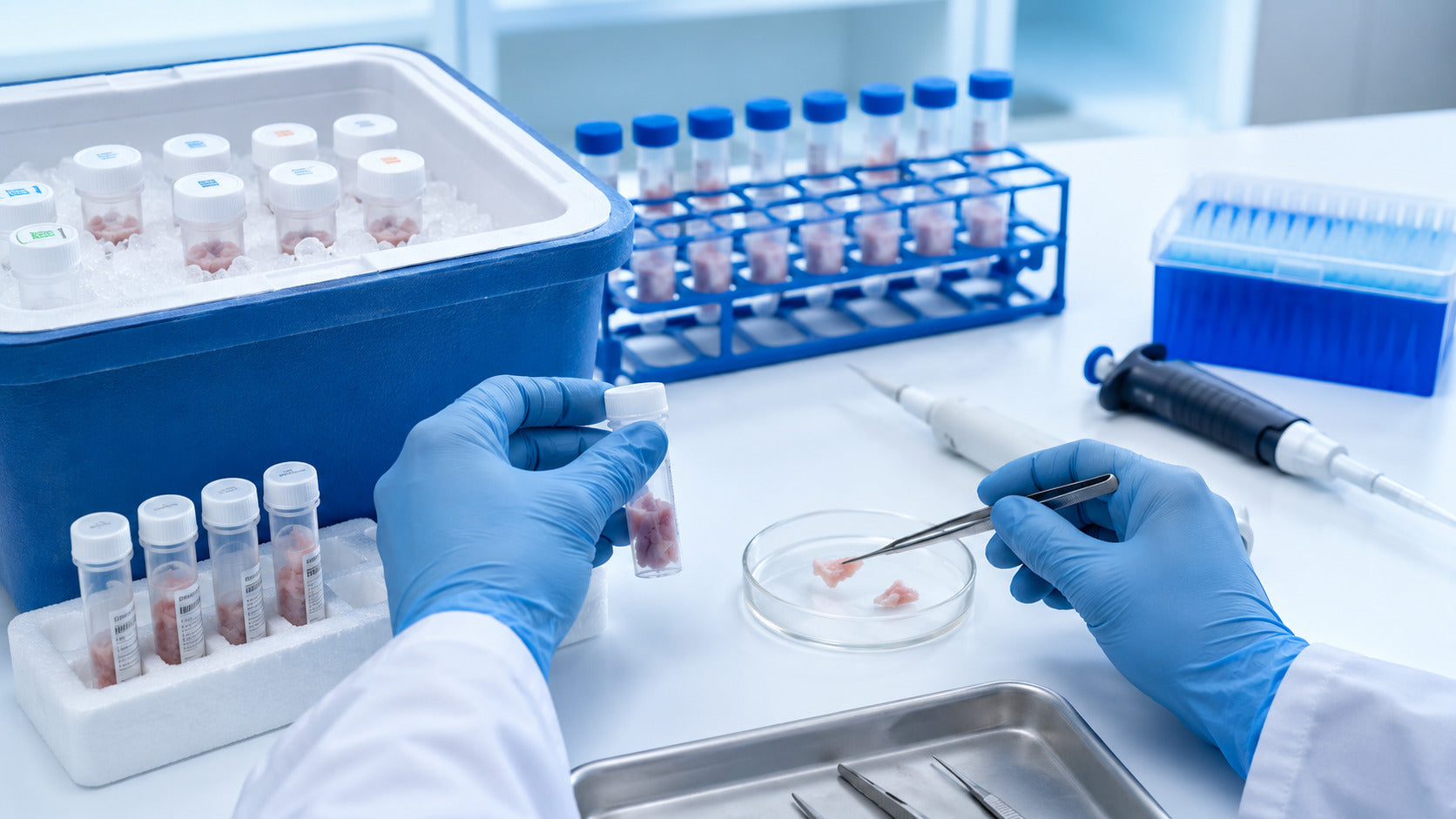
Human tissues should be collected, transported, stored, and prepared according to the intended downstream analysis. For formalin-fixed paraffin-embedded histology, tissues are promptly fixed in 10% neutral buffered formalin before dehydration, clearing, and paraffin infiltration. For flow cytometry, fresh tissue is generally maintained under suitable cold conditions and gently dissociated into a clean single-cell suspension.
To ensure reliable results, human tissue must be processed into a single-cell suspension prior to flow cytometry analysis. The optimal preparation method is determined by tissue type, target cell populations, specific markers, and whether the sample is processed fresh, fixed, frozen, or cryopreserved. A carefully designed workflow protects fragile cells, minimizes aggregates, preserves surface antigens, and enhances reproducibility across experiments.
Scientific Basis and Practical Scope
Research-use human tissue preparation workflows, particularly for applications such as flow cytometry, immunophenotyping, cell sorting, or single-cell analysis. The recommendations are informed by established cytometry literature, specimen-handling principles, and practical considerations from tissue-dissociation workflows. Human tissue preparation requires a tailored approach based on tissue source, marker sensitivity, cell type of interest, storage conditions, and downstream assays. Laboratories should validate transport media, storage duration, dissociation enzymes, digestion temperature, and staining conditions for each specific tissue type and antibody panel prior to implementing workflows in large-scale studies.
The primary objectives are to preserve cell viability, maintain marker expression, minimize debris and aggregates, and produce a clean single-cell suspension suitable for reliable flow cytometry acquisition, thereby supporting the workflow described below.
What Is Human Tissue Handling?
Human tissue handling includes all steps to protect tissue quality from collection through analysis. This covers tissue collection, labelling, transport, storage, dissociation, cleanup, fixation, cryopreservation, and final preparation for flow cytometry. Effective human tissue handling preserves cell viability, surface antigen expression, intracellular markers, cell-type representation, sample cleanliness, and downstream staining quality. Given the limited availability and clinical significance of human tissues, each step must be designed to maintain sample integrity and facilitate subsequent analysis.
Why Human Tissue Preparation Matters for Flow Cytometry
Flow cytometry analyzes individual cells as they pass through the instrument. Consequently, samples must contain well-separated cells with minimal clumping, debris, and dead cells. For solid tissue flow cytometry, tissue preparation is especially important because the tissue must first be converted into an adequate single-cell suspension. Accordingly, tissue dissection, enzymatic digestion, mechanical dissociation, filtration, and washing all influence the final result.
Flow Cytometry Needs a Single-Cell Suspension
A single-cell suspension allows each cell to be stained and analysed individually. If the sample contains aggregates, tissue fragments, or debris, gating becomes harder, and the final data may be less clear.
Cell Viability Improves Data Quality
High cell viability supports cleaner staining and more confident interpretation. Dead cells can bind antibodies non-specifically, release DNA, increase clumping, and create a background signal.
Marker Preservation Supports Accurate Staining
Some markers are sensitive to digestion enzymes, storage time, temperature, and fixation. Therefore, a strong tissue preparation workflow should match the dissociation method to the markers being measured.
Human Tissue Collection Guidelines
Human tissue collection is the first step in sample quality control. The goal is to move tissue from collection to preservation or processing as smoothly as possible, so later handling starts with the best possible material.
Collect Tissue Promptly
Tissue should be placed into the appropriate transport medium or storage condition as soon as possible after collection. Delays can reduce cell viability and alter marker expression.
Use Sterile Tools and Containers
Sterile containers and clean tools help reduce contamination and protect the sample. Containers should be clearly labelled with sample ID, tissue type, collection time, and study information.
Keep Tissue Moist
Fresh tissue should not be allowed to dry. Drying can damage cells and reduce recovery. Tissue should be placed into suitable transport media, buffer, or a validated preservation condition.
Avoid Compression
Tissue should not be crushed or tightly packed in small containers. Mechanical pressure can damage fragile cells and reduce the consistency of dissociation.
Record Sample Metadata
Document tissue source, collection time, transport time, storage condition, temperature, and processing start time. These details help explain variation between samples and improve reproducibility.

Tissue Transport and Storage Before Flow Cytometry
Tissue transport and storage depend on tissue type, study goal, marker panel, and expected time before processing. The best condition should be validated for the specific workflow.
Transport Medium
Common transport options may include culture medium, buffered saline, or a validated tissue preservation solution. The medium should keep tissue moist and be compatible with downstream tissue dissociation and staining.
Transport Temperature
Many fresh-tissue workflows use cold transport to reduce cellular stress and preserve sample quality. However, some markers or functional assays may require specific handling conditions. The receiving laboratory’s SOP should guide the final decision.
Short-Term Fresh Tissue Storage
Fresh tissue may be held briefly in a suitable medium or buffer before dissociation. The storage time should be as short as practical and validated for the tissue type and markers of interest. Fixed or Frozen Tissue Storage. Fixation may be useful when target markers are stable after fixation or when immediate live-cell analysis is not needed. Frozen tissue may be useful for nuclei preparation or other downstream applications. For live-cell flow cytometry, freezing whole tissue should be validated before routine use.e.
Cryopreservation of Human Tissue or Cells
Cryopreservation allows samples to be stored and analysed later. This is useful for multicenter studies, batch staining, and long-term sample banking.
When Cryopreservation Is Useful
Cryopreservation can help when samples arrive at different times or when staining must be performed in batches. It also allows valuable human tissue samples to be preserved for future analysis.
Cryopreserving Dissociated Cells
In many workflows, tissue is first dissociated into cells and then cryopreserved in a validated freezing medium. This can improve consistency compared with freezing unprocessed tissue, depending on the tissue type and assay.
Effects on Viability and Markers
Cryopreservation may affect some cell populations or surface markers. For this reason, thawing workflows should include viability checks and marker validation.
How to Prepare Human Tissue for Flow Cytometry
Preparing human tissue for flow cytometry usually includes trimming, washing, mincing, tissue dissociation, filtering, counting, and staining.
Step 1: Inspect and Trim Tissue
Remove unwanted material such as fat, necrotic regions, large blood clots, or non-target tissue when appropriate. This improves the quality of the starting material.
Step 2: Wash the Tissue
A gentle wash can remove excess blood, transport medium, and loose debris. Blood-rich samples may require red blood cell lysis later in the workflow.
Step 3: Mince Into Small Pieces
Mincing increases surface area and improves tissue dissociation. Pieces should be small and uniform, without aggressively crushing the tissue.
Step 4: Choose the Dissociation Method
The dissociation method depends on tissue density, marker sensitivity, desired cell types, and the downstream assay. Some tissues can be processed mechanically, while dense or fibrotic tissues may need enzymatic digestion.
Step 5: Filter the Suspension
Filtering removes tissue fragments and aggregates. Filter size should match the cell type, sample condition, and flow cytometer requirements.
Step 6: Count Cells and Check Viability
Before staining, measure cell concentration and viability. This helps standardise antibody staining and improves acquisition quality.
Mechanical and Enzymatic Tissue Dissociation
Tissue dissociation is the process of converting solid tissue into individual cells. It may be mechanical, enzymatic, or a combination of both.
Mechanical Tissue Dissociation
Mechanical dissociation uses physical force to separate cells from tissue. Methods may include mincing, teasing, scraping, gentle pipetting, or pressing tissue through a mesh. Mechanical methods can be fast and may reduce changes in enzyme-related markers. They are useful for softer tissues or samples where preservation of surface markers is highly important.
Enzymatic Tissue Dissociation
Enzymatic tissue dissociation uses enzymes to break down the extracellular matrix and release cells. Common enzymes include collagenase, dispase, DNase, trypsin-like enzymes, and tissue-specific enzyme blends. Enzymatic methods can improve yield from solid, dense, or fibrotic tissues. Digestion time, temperature, enzyme concentration, and mixing should be optimised. Over-digestion may affect viability or marker expression, while under-digestion may reduce cell yield.
DNase for Clump Reduction
DNase can help reduce clumping caused by extracellular DNA released from damaged cells. This is useful when samples are sticky, debris-rich, or high in dead cells.
Preparing Single-Cell Suspensions from Body Fluids
Some human samples are already fluid-based, such as fine-needle aspirates, pleural effusions, abdominal fluids, or other body fluids. These may need less tissue dissociation but still require cleanup.
Washing and Debris Removal
Body fluid samples may contain protein, mucus, red blood cells, dead cells, or debris. Washing and filtering can improve sample quality.
Red Blood Cell Lysis
Blood-rich samples may need red blood cell lysis if red cells interfere with staining or acquisition.
Cell Counting and Viability
Cell concentration and viability should be checked before staining to support consistent antibody labelling.
Preparing Intact Nuclei from Human Tissue
In some workflows, intact nuclei are prepared instead of whole cells. This may be useful for frozen tissue, paraffin-embedded tissue, or applications focused on nuclear markers.
Fresh Tissue Nuclei
Fresh tissue can be used for nuclei preparation when whole-cell recovery is not the goal.
Frozen Tissue Nuclei
Frozen tissue can support nuclei-based workflows when live cells are not required. This can be useful for archived tissue studies.
Paraffin-Embedded Tissue Nuclei
Paraffin-embedded tissue may be used for specific nuclei preparation workflows, depending on assay requirements and marker compatibility.
Debris Removal and Sample Cleanup
Debris removal is important because debris can affect gating, increase background, and reduce the quality of flow cytometry data.
Filtration
Cell strainers remove large clumps and tissue fragments. Gentle filtration supports a cleaner suspension.
Dead Cell Removal
Dead cell removal may be useful when viability is low and dead cells interfere with staining or sorting.
Red Blood Cell Removal
Blood-rich human tissue samples may benefit from red blood cell lysis or additional washing.
Density-Based Cleanup
Some samples may benefit from density separation when debris, dead cells, or unwanted fractions are high.
Quality Control Before Flow Cytometry Staining
Quality control should be performed before staining whenever possible.
Cell Viability
Viability dyes help identify dead cells and improve gating. High viability supports cleaner analysis.
Cell Concentration
Standardised cell input improves staining consistency and reduces antibody variability.
Debris and Aggregates
Check the suspension visually or under a microscope. If clumps are present, filter again or optimize dissociation.
Marker Preservation
If a key marker is weak or inconsistent, review digestion conditions, storage time, fixation, and antibody compatibility.
Troubleshooting Table for Human Tissue Preparation
|
Issue |
Possible Cause |
Practical Solution |
|
Low viability |
Long delay, drying, harsh digestion |
Process faster, keep tissue moist, optimize digestion |
|
Low cell yield |
Incomplete dissociation |
Improve mincing, enzyme choice, or incubation time |
|
High debris |
Necrotic tissue or harsh processing |
Trim tissue, reduce force, add cleanup |
|
Cell clumping |
Released DNA or poor filtration |
Use DNase, filter gently, reduce concentration |
|
Weak staining |
Marker affected by digestion or storage |
Adjust enzyme, time, temperature, or fixation |
|
Variable results |
Inconsistent handling |
Standardize SOP and record metadata |
Best Practices for Handling Human Tissue Samples
Good tissue handling is built on consistency. Prepare tools, buffers, media, labels, and documentation before the sample arrives. Keep tissue moist and protected. Minimise the time from collection to processing. Choose tissue dissociation methods based on tissue type and marker sensitivity. Validate storage and cryopreservation conditions before using them in large studies. Human tissue collection and storage guidelines should also include sample acceptance criteria. For example, define acceptable transport time, temperature condition, tissue appearance, minimum viability, and minimum cell recovery.
FireGene Support for Human Tissue Preparation Workflows
FireGene supports tissue sample preparation workflows with research-use solutions for tissue dissociation, sample preservation, cryopreservation, and post-processing cleanup. These workflow tools are useful when researchers need cleaner single-cell suspensions for flow cytometry, cell sorting, single-cell sequencing, or related applications. For human tissue handling, FireGene’s sample preparation resources can help laboratories standardise tissue preparation, improve suspension quality, and support reproducible downstream analysis.
Quick Human Tissue Preparation Checklist
|
Step |
Best Practice |
|
Collection |
Collect promptly and avoid tissue drying |
|
Transport |
Use validated medium and temperature |
|
Storage |
Match storage method to assay goal |
|
Dissociation |
Choose mechanical, enzymatic, or combined method |
|
Cleanup |
Filter, wash, and remove debris when needed |
|
QC |
Check viability, concentration, debris, and clumps |
|
Staining |
Use antibody panel compatible with preparation method |
|
Documentation |
Record source, timing, storage, and processing notes |
FAQs
How do you prepare human tissue for flow cytometry?
Human tissue is prepared by promptly collecting it, keeping it moist, transporting it under validated conditions, mincing it into small pieces, mechanically or enzymatically dissociating it, filtering the suspension, counting cells, assessing viability, and staining with an appropriate antibody panel.
What is the best way to store human tissue before flow cytometry?
The best storage method depends on tissue type, target markers, and assay goal. Fresh tissue is often processed as soon as possible, while fixed, frozen, or cryopreserved samples may be used when validated for the workflow.
Why is single-cell suspension important for flow cytometry?
Flow cytometry analyses individual cells in suspension. A clean single-cell suspension improves staining, gating, acquisition, and data interpretation.
What is tissue dissociation?
Tissue dissociation is the process of breaking solid tissue into individual cells using mechanical force, enzymes, or a combination of both.
What causes low cell viability after tissue preparation?
Low viability can be caused by delayed processing, tissue drying, poor storage, harsh digestion, over-mixing, temperature stress, or repeated freeze-thaw cycles.
How can tissue clumping be reduced?
Clumping can be reduced by gentle dissociation, DNase treatment when appropriate, proper filtration, suitable buffer conditions, and avoiding excessive cell concentration.
Can paraffin-embedded tissue be used for nuclei preparation?
Yes, paraffin-embedded tissue may be used for specific nuclei preparation workflows, depending on assay requirements and marker compatibility.
What are the best practices for handling human tissue samples?
Best practices include prompt collection, suitable transport media, validated storage, gentle dissociation, debris cleanup, viability testing, marker validation, and complete documentation.
Conclusion
Handling, storage, and preparation of human tissues are key steps for successful flow cytometry. A high-quality sample preparation workflow helps preserve cell viability, protect markers, reduce debris, and generate a clean single-cell suspension. By following clear guidelines for human tissue collection and storage, choosing the appropriate tissue dissociation method, validating cryopreservation as needed, and performing QC before staining, researchers can improve the reliability of flow cytometry and make better use of valuable human tissue samples. FireGene supports this positive, quality-focused approach by helping researchers connect tissue handling, sample preparation, and downstream analysis into a more consistent workflow.
Scientific References
- Dressler LG, Visscher D. Handling, storage, and preparation of human tissues. Current Protocols in Cytometry.
- Reichard A, Asosingh K. Best Practices for Preparing a Single Cell Suspension from Solid Tissues for Flow Cytometry. Cytometry Part A.
- Leelatian N, Doxie DB, Greenplate AR, et al. Preparing Viable Single Cells from Human Tissue and Tumours for Cytomic Analysis. Current Protocols in Molecular Biology.
- ISAC educational resources on preparing single-cell suspensions from solid tissues for cytometry workflows.


