
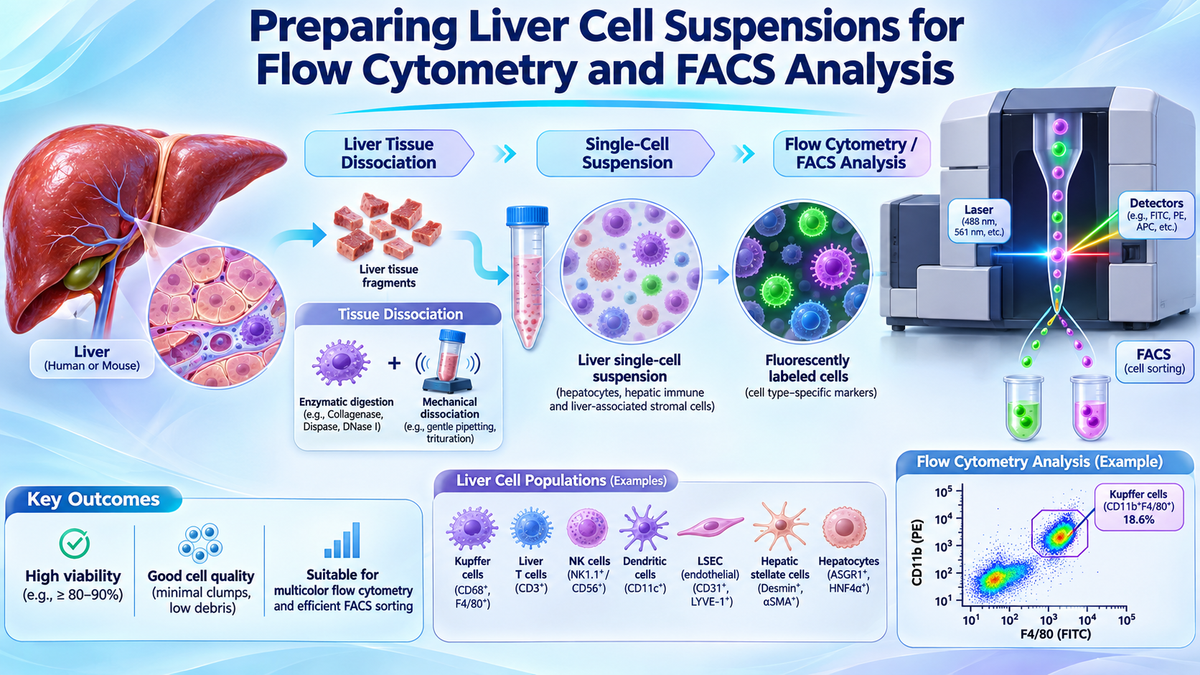
Flow cytometry and fluorescence-activated cell sorting (FACS) are widely used to characterize hepatic cell populations, investigate immune responses, and isolate specific cell types from liver tissue. However, successful analysis depends on obtaining high-quality single-cell suspensions with adequate viability, minimal aggregation, and preserved cell-surface antigens. Liver tissue presents unique challenges because hepatocytes, Kupffer cells, hepatic stellate cells, and liver sinusoidal endothelial cells differ in size, density, and sensitivity to enzymatic digestion. This article discusses the principles of liver cell preparation for flow cytometry and FACS, provides a practical sample preparation workflow, outlines essential quality-control parameters, and explores common troubleshooting strategies for hepatic immunophenotyping and cell sorting.
1. Introduction: The Importance of Liver Cell Preparation for Flow Cytometry
The liver is a highly specialized organ involved in metabolism, detoxification, immune regulation, and tissue regeneration.
It contains multiple cellular populations, including hepatocytes, Kupffer cells, hepatic stellate cells, liver sinusoidal endothelial cells (LSECs), cholangiocytes, and resident or infiltrating immune cells.
These populations contribute to liver homeostasis and participate in pathological processes such as fibrosis, hepatitis, metabolic dysfunction-associated steatotic liver disease (MASLD), and hepatocellular carcinoma (HCC).
Flow cytometry enables researchers to characterize individual cells according to their physical properties and expression of fluorescently labeled molecular markers.
Fluorescence-activated cell sorting (FACS) extends this capability by physically isolating selected cell populations for downstream research.
However, both technologies require appropriately prepared cell suspensions.
High-quality liver cell suspensions are essential for accurate immunophenotyping, reliable cell sorting, and reproducible experimental results.
Poor tissue dissociation can generate aggregates, reduce cell viability, damage surface antigens, and introduce cell-type-specific recovery biases.
Therefore, optimizing liver tissue dissociation and subsequent sample processing is a critical part of successful hepatic flow cytometry.
2. Why Is Liver Tissue Challenging for Flow Cytometry?
2.1 Complex Tissue Architecture
Liver tissue contains organized hepatic plates, sinusoidal networks, and extracellular matrix components.
Collagen, laminin, fibronectin, and other structural proteins maintain tissue integrity but can restrict the release of individual cells during digestion.
Fibrotic or cirrhotic tissue may contain increased extracellular matrix deposition, requiring additional optimization of the dissociation process.
2.2 Differences in Cell Size and Fragility
Hepatocytes are relatively large and mechanically sensitive compared with many non-parenchymal liver cells.
Aggressive pipetting, prolonged digestion, and inappropriate centrifugation can cause cellular damage.
Smaller immune populations may tolerate certain preparation conditions better, potentially resulting in selective recovery.
Consequently, the final cell suspension may not accurately represent the cellular composition of the original liver tissue.
2.3 Cellular Debris and Autofluorescence
Liver tissue may contain damaged cells, tissue fragments, and intracellular components released during dissociation.
These materials can interfere with flow cytometry measurements.
Hepatic cells may also exhibit substantial intrinsic autofluorescence, partly associated with endogenous metabolic molecules and intracellular pigments.
Autofluorescence can complicate the interpretation of weak fluorescence signals, particularly when analyzing low-abundance surface markers.
2.4 Preservation of Cell-Surface Antigens
Enzymatic digestion may alter the accessibility or integrity of certain cell-surface epitopes.
This can influence antibody binding and reduce the accuracy of immunophenotyping.
Researchers should therefore evaluate whether their dissociation procedure is compatible with the markers included in their flow cytometry panel.
3. Step-by-Step Preparation of Liver Cell Suspensions
The following workflow summarizes general approaches used in liver cell preparation. Specific reagent concentrations, incubation conditions, filtration requirements, and centrifugation settings should follow validated protocols appropriate for the tissue source and experimental objective.
Step 1: Liver Tissue Collection and Handling
Fresh liver tissue should be collected and transferred promptly into an appropriate transport or preservation medium.
Processing delays should be minimized because prolonged ischemic exposure may compromise cell viability.
Researchers should record sample characteristics, including tissue source, collection time, sample mass, and preservation conditions.
When appropriate, excess blood, connective tissue, and visibly necrotic regions can be removed before processing.
Consistent sample handling is particularly important when comparing healthy and diseased liver tissues.
Step 2: Tissue Fragmentation
Liver samples are commonly divided into small, relatively uniform fragments to improve enzyme accessibility.
Fragmentation increases the surface area exposed to digestion reagents.
However, excessive mechanical disruption should be avoided because it may damage fragile hepatic populations.
The optimal fragment size depends on tissue condition and the selected dissociation procedure.
Fibrotic tissue may require additional consideration because dense extracellular matrix can reduce digestion efficiency.
Step 3: Enzymatic Liver Tissue Dissociation
Enzymatic digestion is frequently used to release cells from liver tissue.
Collagenase-based systems are commonly employed because they facilitate breakdown of extracellular matrix components.
Depending on the protocol, additional enzymes may be included to improve tissue dissociation or limit aggregation.
Important factors include:
Enzyme activity: Sufficient activity is necessary for effective tissue disruption.
Digestion duration: Prolonged exposure may reduce viability or alter surface antigens.
Temperature: Incubation conditions should follow the validated enzyme protocol.
Mixing intensity: Gentle mixing can improve tissue exposure while minimizing mechanical damage.
The objective is to produce a sufficiently dissociated cell suspension while preserving cellular integrity and relevant surface markers.
Step 4: Gentle Mechanical Dissociation
Following enzymatic digestion, controlled mechanical disruption may help release remaining cells.
Gentle pipetting is commonly used to separate partially digested tissue fragments.
Wide-bore pipette tips may be beneficial when handling large or fragile cells.
Vigorous vortexing and repeated forceful aspiration should generally be avoided.
Excessive mechanical stress can damage cell membranes, increase extracellular DNA release, and promote aggregation.
Step 5: Cell Filtration
The dissociated suspension is usually filtered to remove large tissue fragments and cellular aggregates.
Different filter pore sizes may be used depending on the target population and instrument requirements.
For example, 40 µm and 70 µm strainers are commonly encountered in cell preparation workflows.
However, mature hepatocytes can be relatively large, and overly restrictive filtration may reduce their recovery.
Researchers should therefore select filtration conditions based on target cell size, suspension quality, and downstream requirements.
Step 6: Washing and Cell Resuspension
After filtration, cells are washed to remove residual digestion enzymes and other unwanted components.
Centrifugation conditions should be selected according to the target population.
Hepatocytes and non-parenchymal cells differ in density and sedimentation behavior.
Accordingly, inappropriate centrifugation may cause selective cell loss or damage.
The final suspension should be resuspended in a suitable flow cytometry buffer.
Calcium- and magnesium-free PBS supplemented with an appropriate protein source, such as BSA, is commonly used in certain flow cytometry workflows.
EDTA may help reduce cation-dependent cellular aggregation, but buffer composition must be compatible with the intended assay.
For example, DNase I activity generally requires appropriate divalent cations, so EDTA-containing buffers may be unsuitable during active DNase treatment.
Step 7: Cell Counting and Viability Assessment
Before staining, researchers should assess the quality of the cell suspension.
Important parameters include:
Total cell recovery
Cell viability
Cell concentration
Aggregate frequency
Presence of cellular debris
Representation of target hepatic populations
Trypan blue exclusion and fluorescence-based viability measurements are commonly used.
Cell viability should be evaluated before proceeding with antibody staining or cell sorting.
4. Optimizing Flow Cytometry Staining for Liver Cells
4.1 Selection of Cell-Surface Markers
Flow cytometry panels should be designed according to the research objective and target cell populations.
For hepatic immune profiling, researchers may examine markers associated with leukocytes, macrophages, T cells, B cells, and natural killer cells.
Representative markers include:
|
Cell Population |
Commonly Investigated Markers |
Research Application |
|
Total leukocytes |
CD45 |
Immune-cell identification |
|
T cells |
CD3, CD4, CD8 |
Adaptive immune profiling |
|
B cells |
CD19, CD20 |
B-cell characterization |
|
NK cells |
CD56, CD16 in human samples |
Innate immune research |
|
Monocytes/macrophages |
CD11b, CD14, CD68 |
Myeloid-cell profiling |
|
Kupffer cells |
CLEC4F, TIMD4, VSIG4 |
Liver-resident macrophage studies |
|
Endothelial cells |
CD31, CD146 |
Vascular-cell characterization |
|
Hepatic stellate cells |
Desmin, PDGFRβ, LRAT |
Fibrosis-related research |
Marker availability, specificity, and accessibility vary across species and cellular states.
Some listed markers are intracellular or are not suitable for direct live-cell surface staining. For example, CD68, desmin, and LRAT generally require an appropriate intracellular staining strategy.
Consequently, markers should be selected according to whether the experiment involves live-cell sorting, fixed-cell immunophenotyping, or intracellular protein analysis.
4.2 Viability Staining
Dead cells can exhibit increased nonspecific antibody binding and altered autofluorescence.
Including an appropriate viability dye improves identification of intact, viable populations.
Common options include membrane-impermeant DNA-binding dyes and fixable amine-reactive viability reagents.
The staining approach should be selected according to whether samples will remain viable or undergo fixation and permeabilization.
4.3 Compensation and Fluorescence Controls
Multicolor flow cytometry requires appropriate controls to distinguish overlapping fluorescence signals.
Important controls include unstained samples, single-color controls, and fluorescence-minus-one (FMO) controls when appropriate.
Because liver-derived cells may exhibit substantial autofluorescence, unstained liver-cell controls are particularly valuable.
Spectral flow cytometry can provide additional opportunities to characterize and mathematically separate autofluorescence components.
However, spectral unmixing also requires suitable reference controls and careful validation.
5. Preparing Liver Cells for FACS Sorting
FACS enables the physical isolation of selected hepatic cell populations based on fluorescence characteristics.
Potential applications include isolation of immune-cell subsets, enrichment of specific endothelial populations, and recovery of selected hepatic cells for downstream molecular analysis.
5.1 Maintaining Cell Viability During Sorting
FACS exposes cells to fluidic and mechanical forces.
Fragile liver-derived cells may be particularly sensitive to sorting conditions.
Researchers should coordinate with the flow cytometry facility to select appropriate sample concentration, nozzle diameter, sorting pressure, and collection conditions.
Large hepatocytes may require different sorting settings from smaller lymphocyte populations.
5.2 Preventing Aggregation and Instrument Clogging
Cell aggregates can interfere with sample acquisition, disrupt sorting stability, and reduce recovery.
To minimize aggregation, researchers should:
Prepare sufficiently dissociated cell suspensions.
Use gentle resuspension techniques.
Avoid unnecessarily high cell concentrations.
Consider compatible anti-aggregation strategies.
Filter the suspension immediately before sorting when appropriate.
Filtration must be selected carefully because aggressive removal of large particles may also eliminate target hepatocytes.
5.3 Collection and Post-Sort Recovery
Sorted cells should be collected in an appropriate medium or buffer compatible with downstream applications.
For cell culture experiments, post-sort viability and functional integrity are especially important.
For RNA analysis, minimizing handling delays and maintaining suitable collection conditions can help preserve sample quality.
Post-sort purity and cell recovery should be assessed when sufficient material is available.
6. Common Problems and Troubleshooting Strategies
6.1 Low Cell Recovery
Low yield may result from incomplete digestion, tissue damage, or cell loss during filtration and washing.
Researchers should evaluate the efficiency of each processing step and identify where viable cells are lost.
Increasing digestion intensity should be approached cautiously because it may compromise cellular integrity.
6.2 Reduced Cell Viability
Low viability may reflect prolonged tissue handling, excessive mechanical stress, overdigestion, or inappropriate centrifugation.
Gentle processing and consistent handling conditions can help preserve viable hepatic cells.
6.3 Cell Aggregation
Extracellular DNA released from damaged cells can increase suspension viscosity and promote aggregation.
Potential solutions include reducing mechanical damage, optimizing digestion, and evaluating compatible DNase treatment.
6.4 High Autofluorescence
Autofluorescence may interfere with the detection of weak fluorescence signals.
Researchers should include unstained controls and carefully select fluorochromes.
Where available, spectral flow cytometry and appropriate autofluorescence extraction methods may improve population resolution.
6.5 Poor Marker Detection
Weak or inconsistent marker staining may arise from inappropriate antibody selection, low antigen expression, or enzymatic modification of epitopes.
Researchers should verify antibody performance, review digestion compatibility, and optimize staining conditions.
6.6 Inaccurate Cell Population Representation
Different hepatic populations can be selectively lost during dissociation and separation.
Researchers should avoid assuming that recovered cell proportions accurately represent the original tissue composition.
Complementary methods such as immunohistochemistry, microscopy, or transcriptomic analysis may help validate biological findings.
7. Quick Troubleshooting Table
|
Problem |
Possible Cause |
Recommended Approach |
|
Low cell yield |
Incomplete digestion or processing loss |
Optimize digestion and minimize unnecessary handling |
|
Low viability |
Overdigestion or mechanical stress |
Use gentle manipulation and validated processing conditions |
|
Cell clumping |
Extracellular DNA or residual tissue fragments |
Improve dissociation and consider compatible DNase treatment |
|
High background fluorescence |
Dead cells or intrinsic autofluorescence |
Use viability dyes and appropriate fluorescence controls |
|
Weak antigen staining |
Epitope damage or unsuitable antibody |
Validate marker compatibility with dissociation reagents |
|
Sorter clogging |
Large aggregates or inappropriate concentration |
Optimize sample concentration and filtration |
|
Poor post-sort recovery |
Sorting stress or unsuitable collection conditions |
Adjust sorting parameters and collection medium |
|
Cell-type recovery bias |
Selective loss during dissociation or separation |
Evaluate target population recovery and cell size |
8. Applications of Liver Flow Cytometry and FACS
8.1 Liver Immunology
Flow cytometry allows researchers to investigate resident and infiltrating immune populations in liver tissue.
Applications include profiling T-cell subsets, macrophage populations, NK cells, and inflammatory responses.
FACS can further isolate selected immune populations for gene expression analysis or functional studies.
8.2 Liver Fibrosis Research
Liver fibrosis involves inflammatory signaling, hepatic stellate cell activation, and extracellular matrix accumulation.
Flow cytometry can help characterize immune-cell recruitment and fibrosis-associated cellular changes.
Cell sorting may support downstream characterization of selected stromal and immune populations.
8.3 Hepatocellular Carcinoma Research
The HCC tumor microenvironment contains heterogeneous immune and stromal populations.
Flow cytometry can investigate tumor-infiltrating lymphocytes, macrophages, and other immune cells.
FACS may be used to isolate selected populations for molecular or functional analysis.
8.4 Liver Regeneration and Injury
Flow cytometry supports investigations into cellular responses following liver injury and regeneration.
Researchers can examine changes in immune-cell populations, cellular activation, and selected hepatic cell subsets.
8.5 Integration with Single-Cell Transcriptomics
Flow cytometry and FACS can complement single-cell RNA sequencing.
For example, FACS may be used to enrich selected immune populations before transcriptomic analysis.
However, enrichment introduces intentional changes to cell-type proportions and should be considered during downstream interpretation.
9. Supporting Hepatic Cell Preparation with FireGene
Reliable flow cytometry begins with an appropriately prepared single-cell suspension.
The FireGene Liver Dissociation Kit for Hepatic Cell Profiling (FG-BA3323) is designed to support enzymatic dissociation of liver tissue for downstream cellular research.
The kit provides a liver-focused tissue preparation option that may be incorporated into workflows involving hepatic cell characterization.
Potential applications include:
Liver single-cell suspension preparation
Hepatic immune-cell profiling
Flow cytometry sample preparation
FACS-based cell enrichment
Liver fibrosis and inflammation research
Single-cell transcriptomic studies
Using a standardized dissociation reagent system may help researchers establish consistent sample preparation procedures.
Nevertheless, the suitability of the resulting suspension for flow cytometry or FACS depends on tissue condition, cell viability, antigen preservation, filtration, and downstream handling.
Researchers should follow the current FireGene product instructions and validate compatibility with their specific antibody panels and sorting requirements.
The kit supports liver tissue dissociation and does not replace antibody staining reagents, cell sorters, or cell-type-specific purification protocols.
Explore the FireGene Liver Dissociation Kit for Hepatic Cell Profiling:
https://firegene.com/products/liver-dissociation-kit-fg-ba3323
10. Frequently Asked Questions
How do you prepare liver cells for flow cytometry?
Liver tissue is typically processed using enzymatic and controlled mechanical dissociation, followed by filtration, washing, cell counting, and appropriate antibody staining. The workflow should be optimized according to the target population.
What causes cell clumping during liver FACS preparation?
Clumping can result from incomplete tissue dissociation, extracellular DNA released from damaged cells, and inappropriate suspension conditions.
Can hepatocytes be analyzed using flow cytometry?
Yes. However, hepatocytes are relatively large and fragile, and instrument settings, filtration, antibody selection, and sample preparation must be adapted accordingly.
Which markers are used to identify Kupffer cells?
Markers such as CLEC4F, TIMD4, and VSIG4 are frequently investigated in Kupffer cell research. Marker specificity depends on species, disease context, and the relevant macrophage populations.
How can autofluorescence be reduced in liver flow cytometry?
Appropriate controls, fluorochrome selection, viability gating, and spectral autofluorescence extraction may improve the distinction between genuine staining and background fluorescence.
What is the difference between flow cytometry and FACS?
Flow cytometry measures cellular characteristics using light scattering and fluorescence. FACS additionally isolates selected cells into separate collection vessels for further research.
11. Conclusion
Preparing high-quality liver cell suspensions is fundamental to successful flow cytometry and FACS analysis.
Effective workflows require an appropriate balance between tissue dissociation efficiency, cell viability, surface antigen preservation, and cellular representation.
Special attention should be given to the structural complexity of liver tissue, hepatocyte fragility, aggregation, and intrinsic autofluorescence.
Optimized enzymatic digestion, gentle sample handling, suitable filtration, and rigorous quality control can improve the consistency of hepatic cellular analysis.
The FireGene Liver Dissociation Kit for Hepatic Cell Profiling provides a liver-focused dissociation reagent option that can be incorporated into validated sample preparation workflows.
Ultimately, selecting the appropriate preparation strategy according to the target cell populations and downstream research objectives is essential for generating reliable flow cytometry and FACS results.
For Research Use Only. Not for diagnostic or therapeutic use.


