
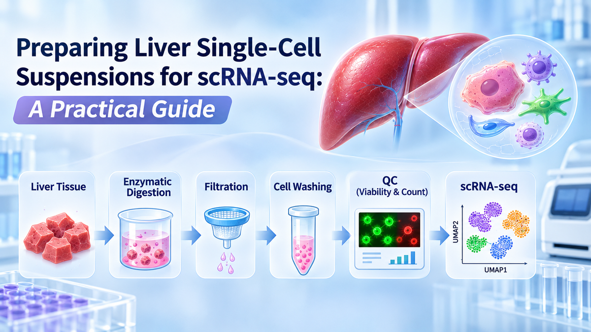
Single-cell RNA sequencing (scRNA-seq) has transformed liver research by enabling high-resolution characterization of hepatic cell populations, transcriptional states, and disease-associated molecular pathways. However, obtaining reliable scRNA-seq data depends heavily on the quality of the initial single-cell suspension. Liver tissue presents particular challenges because hepatocytes are relatively large and fragile, while non-parenchymal cells differ considerably in size, density, and sensitivity to tissue dissociation. Inadequate sample preparation can cause cell loss, aggregation, ambient RNA contamination, and biased cell-type representation. This article outlines practical strategies for preparing liver single-cell suspensions, discusses essential quality-control parameters, and highlights approaches for improving sample consistency in hepatic transcriptomics research.
1. Introduction: The Importance of Sample Preparation in Liver scRNA-seq
Single-cell RNA sequencing has become an important technology for investigating liver cellular heterogeneity and disease-associated transcriptional changes.
Unlike conventional bulk RNA sequencing, which measures average gene expression across mixed cell populations, scRNA-seq enables researchers to characterize individual cells and identify transcriptionally distinct subpopulations.
The liver contains multiple cellular populations, including:
Hepatocytes
Kupffer cells and other macrophages
Hepatic stellate cells
Liver sinusoidal endothelial cells (LSECs)
Cholangiocytes
Resident and infiltrating immune cells
These populations participate in metabolism, immune regulation, extracellular matrix remodeling, bile secretion, and tissue regeneration.
Single-cell approaches have been applied to liver fibrosis, metabolic dysfunction-associated steatotic liver disease (MASLD), hepatocellular carcinoma, and inflammatory liver disorders.
However, high-quality liver scRNA-seq begins with successful tissue dissociation and single-cell suspension preparation.
Poor-quality suspensions may introduce technical artifacts that affect cellular recovery, sequencing efficiency, and biological interpretation.
Consequently, optimizing the preparation workflow is essential for obtaining reliable hepatic single-cell transcriptomic data.
2. Why Is Liver Single-Cell Preparation Challenging?
2.1 Hepatocyte Size and Fragility
Hepatocytes are relatively large cells with specialized structural and metabolic characteristics.
They can be particularly sensitive to mechanical stress, prolonged enzymatic digestion, and inappropriate centrifugation.
Excessive processing may lead to membrane damage, cellular rupture, and reduced viable hepatocyte recovery.
Because hepatocytes contain abundant RNA, cell rupture may also contribute substantially to ambient RNA contamination.
2.2 Complex Extracellular Matrix
Liver tissue contains extracellular matrix components, including collagen, laminin, and fibronectin.
These molecules help maintain hepatic architecture but can restrict the release of individual cells during tissue dissociation.
Fibrotic and cirrhotic samples present additional challenges because increased extracellular matrix deposition may require modified digestion strategies.
2.3 Differences Among Hepatic Cell Populations
Hepatocytes, macrophages, stellate cells, and endothelial cells differ in size, density, adhesion, and susceptibility to enzymatic treatment.
Therefore, a dissociation method that efficiently recovers one population may cause selective loss of another.
This issue is especially important when the research objective is to characterize the complete hepatic cellular landscape.
2.4 Dissociation-Induced Transcriptional Changes
Tissue collection and dissociation can induce cellular stress responses.
Previous studies have demonstrated that tissue processing may alter the expression of immediate-early genes and stress-associated transcriptional programs.
These changes can complicate comparisons between experimental groups.
Researchers should therefore consider both cell viability and preservation of biological transcriptional states during sample preparation.
3. Step-by-Step Workflow for Liver Single-Cell Suspension Preparation
The following workflow describes general principles used in hepatic single-cell preparation. Exact reagent concentrations, incubation conditions, centrifugation settings, and filtration requirements should be established using validated tissue-specific protocols and the manufacturer's instructions.
Step 1: Liver Tissue Collection and Initial Handling
Fresh liver tissue should be collected and transferred promptly into a suitable transport or preservation medium.
Delays between tissue collection and processing can contribute to ischemic stress, reduced cell viability, and RNA degradation.
Researchers should document important sample characteristics, including tissue source, sample mass, collection time, preservation conditions, and visible pathological changes.
When appropriate, unwanted connective tissue, blood, and visibly necrotic regions may be removed before dissociation.
Consistent sample handling is particularly important when comparing normal and diseased liver tissues.
Step 2: Tissue Fragmentation
Liver tissue is generally divided into small, relatively uniform fragments to facilitate enzymatic access.
Fragmentation increases the surface area exposed to digestion reagents and can improve processing consistency.
However, excessive mechanical disruption should be avoided because it may damage fragile hepatocytes.
The optimal fragment size depends on tissue properties, sample quantity, and the dissociation protocol.
Fibrotic samples may require additional attention because dense extracellular matrix can reduce digestion efficiency.
Step 3: Enzymatic Liver Tissue Dissociation
Enzymatic digestion is a central component of many liver single-cell isolation workflows.
Collagenase-based systems are widely used to disrupt extracellular matrix components and facilitate hepatic cell release.
Depending on the method, other enzymes may be incorporated to improve tissue processing or reduce aggregation.
Important parameters include:
Enzyme activity: Appropriate enzymatic activity is required for efficient tissue dissociation.
Digestion duration: Insufficient digestion may leave cell aggregates, whereas prolonged exposure can reduce cell viability.
Incubation temperature: Temperature should be controlled according to the enzyme formulation and validated protocol.
Mixing conditions: Gentle mixing may improve tissue exposure to digestion reagents while limiting mechanical damage.
The goal is to achieve sufficient tissue disruption without unnecessarily compromising cellular integrity.
Step 4: Gentle Mechanical Dissociation
Following enzymatic treatment, controlled mechanical dissociation may help release additional cells.
Gentle pipetting can assist in separating partially digested tissue fragments.
Wide-bore pipette tips may be beneficial for fragile or relatively large cells.
Aggressive pipetting, repeated forceful aspiration, and unnecessary vortexing should generally be avoided.
Excessive mechanical stress can rupture hepatocytes, increase extracellular DNA release, and generate debris.
Step 5: Cell Filtration and Aggregate Removal
After dissociation, the suspension is commonly filtered to remove large tissue fragments and cell aggregates.
Cell strainers of different pore sizes may be selected depending on the target population and downstream application.
For example, 40 µm and 70 µm filters are used in various single-cell workflows.
However, mature hepatocytes can be relatively large, and restrictive filtration may reduce their recovery.
Researchers should therefore balance aggregate removal against cell-type-specific losses.
For droplet-based scRNA-seq, minimizing aggregates is particularly important because cell clusters can interfere with microfluidic processing and increase multiplet formation.
Step 6: Cell Washing and Suspension Preparation
Following filtration, cells are typically washed to remove residual digestion reagents, cellular debris, and soluble contaminants.
Centrifugation conditions should be selected according to the intended cell populations.
Large hepatocytes and smaller non-parenchymal cells differ in sedimentation behavior, making separation conditions an important source of potential recovery bias.
Repeated washing may improve suspension cleanliness but can also increase cell loss.
The final cell suspension should be prepared using a buffer compatible with the selected sequencing platform.
For example, PBS supplemented with 0.04% BSA is commonly recommended for certain 10x Genomics fresh-cell workflows.
However, buffer compatibility must be verified for the specific assay, particularly when using different library preparation chemistries.
Step 7: Cell Counting and Viability Assessment
Before sequencing, cell concentration and viability should be evaluated using a suitable counting method.
Common approaches include trypan blue exclusion and fluorescence-based live/dead staining.
Important parameters include:
Total recovered cell number
Viable cell percentage
Cell concentration
Aggregate frequency
Residual debris
Cell size distribution
For many conventional droplet-based fresh-cell workflows, viability of approximately 90% or higher is desirable, although acceptable thresholds depend on the sequencing platform and application.
Viability alone is not sufficient to establish sample quality. Aggregate content, cell-type representation, and extracellular RNA contamination should also be considered.
4. Key Quality-Control Parameters for Liver scRNA-seq
|
Quality Parameter |
Why It Matters |
Recommended Consideration |
|
Cell viability |
Dead cells may release RNA and increase background contamination |
Aim for high viability according to platform requirements |
|
Cell concentration |
Influences capture efficiency and multiplet risk |
Follow assay-specific loading recommendations |
|
Cell aggregation |
May cause microfluidic problems and multiplets |
Minimize visible clusters before loading |
|
Cellular debris |
Can interfere with counting and sample processing |
Use appropriate cleanup when necessary |
|
Cell-type representation |
Determines the biological populations available for analysis |
Evaluate potential enrichment or depletion bias |
|
RNA integrity |
Supports reliable transcriptomic measurements |
Minimize processing delays and cellular damage |
|
Suspension buffer |
May affect cell stability and assay chemistry |
Use a validated platform-compatible buffer |
Quality-control requirements should be established before initiating tissue collection.
This allows researchers to design sample preparation strategies appropriate for the intended sequencing platform.
5. Common Problems and Troubleshooting Strategies
5.1 Low Cell Yield
Low recovery may result from incomplete enzymatic digestion, poor tissue quality, or cell loss during washing and filtration.
Researchers should evaluate digestion efficiency, tissue fragment size, and cell recovery at individual processing stages.
Increasing digestion intensity should be approached cautiously because improved tissue disruption does not necessarily translate into higher viable cell recovery.
5.2 Reduced Cell Viability
Low viability may reflect prolonged tissue handling, overdigestion, excessive mechanical stress, or inappropriate centrifugation.
Optimizing processing duration and using gentle manipulation techniques can help preserve fragile hepatic cells.
Cell viability should be assessed at consistent workflow checkpoints to identify where damage occurs.
5.3 Cell Clumping and Aggregation
Cell aggregates may develop because of incomplete tissue dissociation or extracellular DNA released from damaged cells.
Potential strategies include improving digestion consistency, minimizing cellular damage, and considering protocol-compatible DNase treatment.
Any residual DNase or other additives must be compatible with the final sequencing chemistry.
5.4 Excessive Ambient RNA
Ambient RNA originates partly from cells that rupture during preparation.
This contamination can produce misleading gene expression signals, particularly when highly expressed hepatocyte transcripts are detected in unrelated cell populations.
Reducing cell damage and removing problematic debris can help limit contamination.
Computational methods such as SoupX may assist with downstream correction, but they do not replace appropriate sample preparation.
5.5 Underrepresentation of Hepatocytes
Some liver scRNA-seq datasets contain fewer hepatocytes than expected from the original tissue composition.
Possible explanations include hepatocyte fragility, size-dependent filtration losses, and selective recovery during centrifugation or sorting.
Researchers should evaluate whether their isolation method adequately represents the populations relevant to their biological questions.
6. Preserving Hepatic Cell Diversity During scRNA-seq Preparation
One of the major objectives of hepatic single-cell profiling is to characterize interactions among different liver cell populations.
However, dissociation and separation procedures can introduce substantial cell-type bias.
For example, hepatocytes may be disproportionately affected by processing stress, whereas some immune populations may survive dissociation more efficiently.
Appropriate cell identity markers can help researchers assess recovered populations.
|
Liver Cell Type |
Representative Markers |
Research Applications |
|
Hepatocytes |
ALB, HNF4A, CYP3A4 |
Metabolism, hepatotoxicity |
|
Kupffer cells |
CD68, CLEC4F, TIMD4 |
Liver immunity, inflammation |
|
Hepatic stellate cells |
LRAT, DES, RGS5 |
Fibrosis, extracellular matrix remodeling |
|
LSECs |
STAB2, LYVE1, CLEC4G |
Sinusoidal biology, vascular signaling |
|
Cholangiocytes |
KRT7, KRT19, EPCAM |
Biliary biology, cholangiopathies |
Marker specificity varies across species, disease states, and cellular activation conditions.
Importantly, cell-type proportions measured by scRNA-seq should not automatically be interpreted as the true cellular composition of the original tissue.
Orthogonal methods, including histology, immunostaining, and flow cytometry, can help validate cellular representation.
7. scRNA-seq vs. snRNA-seq in Liver Research
Single-nucleus RNA sequencing (snRNA-seq) provides an alternative when intact-cell isolation is difficult.
Unlike scRNA-seq, which profiles RNA from individual cells, snRNA-seq primarily captures nuclear transcripts.
This approach may be useful for frozen samples and tissues containing fragile or difficult-to-dissociate cells.
Comparative liver studies have demonstrated that scRNA-seq and snRNA-seq can recover different proportions of hepatic cell populations.
In some settings, snRNA-seq provides improved representation of hepatocytes and certain mesenchymal populations, whereas scRNA-seq may be advantageous for studying selected immune populations.
However, snRNA-seq and scRNA-seq measure different RNA compartments and are not interchangeable.
Researchers should select the method based on sample availability, target cell populations, and experimental objectives.
8. Supporting Liver Single-Cell Preparation with FireGene
The FireGene Liver Dissociation Kit for Hepatic Cell Profiling (FG-BA3323) is designed to support the enzymatic dissociation of liver tissue into single-cell suspensions for downstream research.
The kit provides a liver-focused reagent system intended to facilitate the preparation of hepatic cells, including hepatocytes and non-parenchymal populations.
Potential research applications include:
Single-cell RNA sequencing: Preparation of liver-derived cell suspensions for investigating cellular heterogeneity and transcriptional regulation.
Liver fibrosis research: Characterization of hepatic stellate cells, inflammatory populations, and fibrosis-associated cellular changes.
Hepatocellular carcinoma research: Investigation of liver tumor-associated cells and their surrounding microenvironment.
Hepatic immunology: Preparation of cells for studying Kupffer cells and other liver-associated immune populations.
Drug metabolism and hepatotoxicity: Generation of liver-derived cell suspensions for subsequent cellular characterization and experimental analysis.
By incorporating a standardized liver dissociation reagent system into the workflow, researchers can reduce variability associated with reagent preparation.
However, performance depends on tissue characteristics, sample handling, and downstream processing. Researchers should follow the current product instructions and validate cell yield, viability, and compatibility with the selected sequencing platform.
Explore the FireGene Liver Dissociation Kit for Hepatic Cell Profiling:
https://firegene.com/products/liver-dissociation-kit-fg-ba3323
9. Frequently Asked Questions
What is the ideal viability for liver single-cell RNA sequencing?
High viability is desirable, and approximately 90% or higher is commonly recommended for conventional fresh-cell droplet workflows. However, acceptable thresholds depend on the sequencing platform and experimental design.
How can I reduce hepatocyte loss during sample preparation?
Use gentle handling, avoid excessive digestion, and evaluate filtration and centrifugation conditions that may selectively remove large or fragile cells.
Can frozen liver tissue be used for scRNA-seq?
Frozen tissue generally presents challenges for conventional live-cell scRNA-seq because intact viable cells may not be recoverable. Single-nucleus RNA sequencing is often considered for such samples.
Why is cell aggregation problematic for scRNA-seq?
Aggregates can interfere with microfluidic processing, increase multiplet formation, and compromise cell counting accuracy.
Is liver tissue dissociation sufficient to obtain purified hepatocytes?
No. Tissue dissociation generates a heterogeneous cell suspension. Additional separation or enrichment steps may be required to obtain purified hepatocyte populations.
10. Conclusion
Preparing high-quality liver single-cell suspensions is a critical prerequisite for reliable hepatic scRNA-seq analysis.
Successful sample preparation requires careful coordination of tissue collection, enzymatic digestion, mechanical manipulation, filtration, washing, and quality assessment.
Because different hepatic populations vary in physical characteristics and sensitivity to processing, researchers must balance cell recovery, viability, and cellular representation.
The FireGene Liver Dissociation Kit for Hepatic Cell Profiling provides a liver-oriented enzymatic dissociation option that can be incorporated into optimized hepatic single-cell preparation workflows.
Ultimately, standardized handling, application-specific quality control, and thoughtful interpretation of cell-type recovery are essential for generating meaningful liver single-cell transcriptomic data.
For Research Use Only. Not for diagnostic or therapeutic use.


