
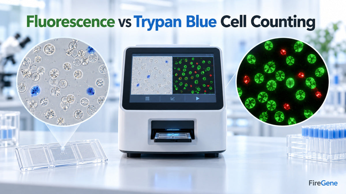
Fluorescence vs Trypan Blue cell counting comes down to sample complexity: Trypan Blue is a practical choice for routine cell culture, while AO/PI fluorescence can provide clearer live/dead cell identification in primary, debris-rich, or heterogeneous samples. Automated cell counting helps researchers measure cell concentration and cell viability with greater speed and consistency than manual counting. Both Trypan Blue staining and fluorescence assays can support dependable results when the method, instrument settings, and sample-preparation workflow are matched to the cell type.
The best method is therefore not simply the one with the most advanced optics. It is the method that provides a representative count for the specific cell sample. Cell size, debris, aggregation, sample concentration, and membrane integrity all influence the final viability assessment. For tissue-derived samples, thoughtful preparation before counting can be equally important. FireGene provides solutions for tissue dissociation, dead-cell removal, debris cleanup, and aggregation control, helping researchers prepare cleaner single-cell suspensions for counting and downstream analysis.
Related Products for Cell Sample Preparation
|
Workflow need |
FireGene solution |
How it supports the workflow |
|
Preparing cells from tissue |
Supports efficient preparation of single-cell suspensions from multiple tissue types |
|
|
Removing unwanted dead cells |
Enriches viable cells before counting, flow cytometry, culture, or sequencing |
|
|
Removing dead cells and tissue debris |
Produces a cleaner suspension for improved sample quality |
|
|
Supporting uniform suspensions |
Helps reduce cell aggregation during sample handling |
These products support the preparation stage before automated cell counting. They are not cell-counting dyes or automated counters.
What Is Automated Cell Counting?
Automated cell counting uses digital imaging and analysis software to identify cells within a defined sample volume. The instrument then calculates cell concentration and, when a viability dye is used, estimates the proportion of live and dead cells.
A typical automated cell counting workflow includes:
- Preparing a uniform cell suspension
- Mixing the sample with a compatible viability dye
- Loading the counting chamber or measurement surface
- Capturing brightfield or fluorescence images
- Identifying and classifying cells
- Reporting total concentration, viable concentration and percentage viability
Automation supports a standardized workflow and reduces the operator-to-operator variation associated with manual hemocytometer counting. However, the quality of the result still depends on representative sampling, accurate pipetting, proper mixing, suitable cell-detection settings, and clean starting material.
How Does Trypan Blue Cell Counting Work?
Trypan Blue staining is based on membrane integrity. Live cells with intact membranes exclude the dye and remain relatively clear, while cells with compromised membranes take up the dye and appear blue. An automated brightfield cell counter captures images of the stained sample and classifies objects according to parameters such as size, shape, brightness, and dye intensity. It then calculates the number of live cells, dead cells, total cells, and overall cell viability.
Benefits of Trypan Blue Staining
Trypan Blue automated cell counting remains popular because it is:
- Simple to integrate into routine laboratory workflows
- Compatible with many brightfield cell counters
- Familiar to researchers working with established cell lines
- Suitable for rapid cell concentration and viability checks
- Cost-effective for regular cell culture monitoring
For clean, well-dispersed cell samples with clearly defined cell boundaries, Trypan Blue can provide fast and useful viability information.
Trypan Blue Counting Considerations
Accurate classification depends on the instrument distinguishing cells from debris and other particles visible in brightfield images. Samples containing tissue fragments, red blood cells, apoptotic bodies, or irregularly shaped cells may require optimized detection settings. Timing also deserves attention. Researchers can standardize the interval between dye mixing and measurement so that every sample is analyzed under comparable conditions. Consistent mixing ratios and careful pipetting further strengthen comparability between experiments.

How Does Fluorescence Cell Counting Work?
Fluorescence cell counting uses fluorescent dyes and optical filters to identify cells according to their staining characteristics. A commonly used approach combines acridine orange and propidium iodide, often called an AO/PI assay.
What Does Acridine Orange Do?
Acridine orange is a nucleic-acid-binding fluorescent dye that enters both live and dead nucleated cells. Under suitable fluorescence optics, it provides a signal that helps the software recognize nucleated cells within the sample. This nucleic-acid-based detection can be especially valuable when the suspension contains noncellular tissue debris. Many debris particles do not produce the same nucleic-acid-associated signal as intact nucleated cells, helping the counter separate cellular events from background material.
What Does Propidium Iodide Do?
Propidium iodide is generally excluded by cells with intact membranes and enters cells with compromised membranes. It therefore provides a fluorescence signal associated with nonviable cells. In a dual-stain AO/PI assay, cells displaying the acridine orange signal without propidium iodide are classified as viable, while propidium-iodide-positive cells are classified as nonviable. The exact displayed colors and classification rules depend on the counter and software.
Benefits of Fluorescence Automated Cell Counting
Fluorescence cell counting is particularly useful for:
- Primary cells isolated from tissues
- Heterogeneous cell populations
- Samples containing substantial debris
- Low-concentration cell suspensions
- Small or irregularly shaped cells
- Workflows requiring confident live/dead classification
- Samples being prepared for flow cytometry or single-cell sequencing
Fluorescence detection can add specificity because classification is based on dye signals associated with nucleated cells and membrane integrity rather than brightfield appearance alone.
Fluorescence vs Trypan Blue Cell Counting
|
Comparison factor |
Trypan Blue counting |
AO/PI fluorescence counting |
|
Detection approach |
Brightfield dye exclusion |
Fluorescent nucleic-acid and membrane-integrity staining |
|
Live-cell identification |
Cells exclude Trypan Blue |
AO-positive and PI-negative classification |
|
Dead-cell identification |
Cells appear blue |
PI-positive fluorescence |
|
Routine cell culture |
Highly practical |
Also suitable |
|
Primary cells |
Effective with optimized settings |
Often useful for complex samples |
|
Debris-rich samples |
Benefits from sample cleanup |
Fluorescence can improve cell-to-debris discrimination |
|
Instrument requirement |
Brightfield-compatible counter |
Fluorescence-enabled counter |
|
Workflow complexity |
Simple |
Requires compatible dyes and optical channels |
|
Best use |
Clean, uniform suspensions |
Complex, heterogeneous or tissue-derived suspensions |
Neither method automatically guarantees a representative result. A clean, single-cell suspension, standardized staining procedure, suitable concentration range, and consistent analysis settings help both methods perform effectively.
Which Cell Counting Method Should You Choose?
Routine Cell Culture Samples
Trypan Blue is a convenient choice for established cell lines that produce uniform, relatively clean suspensions. It supports regular culture maintenance, seeding calculations, passage planning, and routine viability monitoring. Fluorescence can also be selected when the laboratory already uses a fluorescence-enabled counter or wants the same measurement approach across multiple sample types.
Primary Cells and Tissue-Derived Samples
Primary cells often vary in size, shape, granularity, and membrane condition. Tissue dissociation can also generate extracellular material and cellular fragments. AO/PI fluorescence cell counting may offer clearer cellular identification in these complex samples. The decision should be confirmed with the actual tissue type. Running matched aliquots of the same suspension with both methods provides a practical way to compare total concentration, viable concentration, and repeatability.
Samples Containing Debris
Samples with visible debris benefit from a two-part strategy: suitable detection technology and effective sample cleanup. Fluorescence may help distinguish nucleated cells from noncellular material, while debris removal improves the quality of the suspension presented to the instrument. The FireGene Dead Cell Debris Removal Kit uses a density-based workflow to remove dead cells, apoptotic bodies, and unwanted debris from single-cell suspensions. This preparation can support clearer cell samples for counting and subsequent applications.
Samples with Cell Aggregates
Aggregates can contain multiple cells but appear as a single large event, influencing both total cell concentration and viability measurements. Gentle resuspension and aggregation control support a more uniform sample. The FireGene Anti-Clumping Agent is designed to help maintain single-cell suspensions during sample processing. Researchers should confirm compatibility with their cell type and downstream application when integrating any preparation reagent.
Why Sample Preparation Matters Before Cell Counting
Many comparisons focus only on dyes and instruments. A valuable additional question is whether the counted aliquot accurately represents the full sample.
Dead-Cell Content
Dead cells can release intracellular material and contribute to background in cell-based workflows. When higher viable-cell enrichment is desired, the FireGene Dead Cell Removal Solution provides a density-based separation workflow for mammalian tissue-derived single-cell suspensions. After cleanup, researchers can recount the sample and record both viable-cell recovery and percentage viability. This gives a more complete view than percentage viability alone.
Debris and Background Material
Tissue fragments and cellular material can influence object recognition, particularly in brightfield analysis. A dedicated cleanup step helps produce a clearer suspension and can support consistent gating, counting, staining, and downstream processing.
Representative Sampling
Cells naturally settle over time. Gently mixing the suspension immediately before collecting the counting aliquot helps ensure that the measured portion represents the entire sample; consistent pipetting technique and chamber loading further support repeatable results.

A Practical Workflow for Consistent Cell Counting
- Prepare the suspension: Use a tissue-appropriate dissociation method to release cells efficiently.
- Inspect sample quality: Check for aggregates, tissue fragments and visible background material.
- Apply cleanup when beneficial: Remove dead cells or debris according to the sample’s needs.
- Create a uniform suspension: Mix gently and use an anti-clumping approach when appropriate.
- Select the counting method: Choose Trypan Blue for straightforward brightfield counting or AO/PI fluorescence for added cellular specificity.
- Standardize staining: Keep dye ratio, incubation time and temperature consistent.
- Count technical replicates: Repeat measurements to assess sample uniformity.
- Record complete results: Document total concentration, viable concentration, viability percentage, dilution factor, and instrument settings.
- Proceed efficiently: Use the measured viable-cell concentration to normalize downstream input.
This workflow connects cell counting with sample preparation instead of treating the count as an isolated step.

Frequently Asked Questions
Is fluorescence cell counting better than Trypan Blue?
Fluorescence counting is often advantageous for debris-rich, heterogeneous, or primary-cell samples. Trypan Blue remains a practical option for clean and uniform cell culture suspensions. The best method is the one validated for the specific sample.
What is the difference between acridine orange and propidium iodide?
Acridine orange labels nucleated cells, while propidium iodide identifies cells with compromised membranes. Together, AO/PI staining supports live/dead cell classification by fluorescence.
Can Trypan Blue be used for primary cells?
Yes. Trypan Blue can be used with primary cells when the counter settings, staining time, size range, and sample preparation are optimized for that population.
Does debris affect automated cell counting?
Debris can influence object classification, especially when particles resemble cells in size or appearance. Fluorescence-based detection and appropriate sample cleanup can support clearer discrimination.
Should dead cells be removed before counting?
An initial count can characterize the starting suspension. When enrichment is appropriate, dead-cell removal can then be performed, followed by a second count to measure viable-cell recovery and confirm the quality of the prepared sample.
What is more important: viability percentage or viable-cell concentration?
Both are valuable. Viability percentage shows the proportion of live cells, while viable-cell concentration indicates how many usable cells are available per unit volume. Recording both supports better downstream planning.
Conclusion
Fluorescence and Trypan Blue automated cell counting are both valuable viability assessment methods. Trypan Blue provides a simple solution for many routine cultures, while AO/PI fluorescence supports confident identification in complex cell samples.
Reliable results begin with a representative, well-prepared suspension. FireGene’s tissue dissociation, dead-cell removal, debris cleanup, and anti-clumping solutions help researchers prepare high-quality samples before counting, flow cytometry, cell culture, and single-cell sequencing. Explore FireGene single-cell sample-preparation solutions or contact the FireGene team to discuss a workflow matched to your tissue type and downstream application.


