
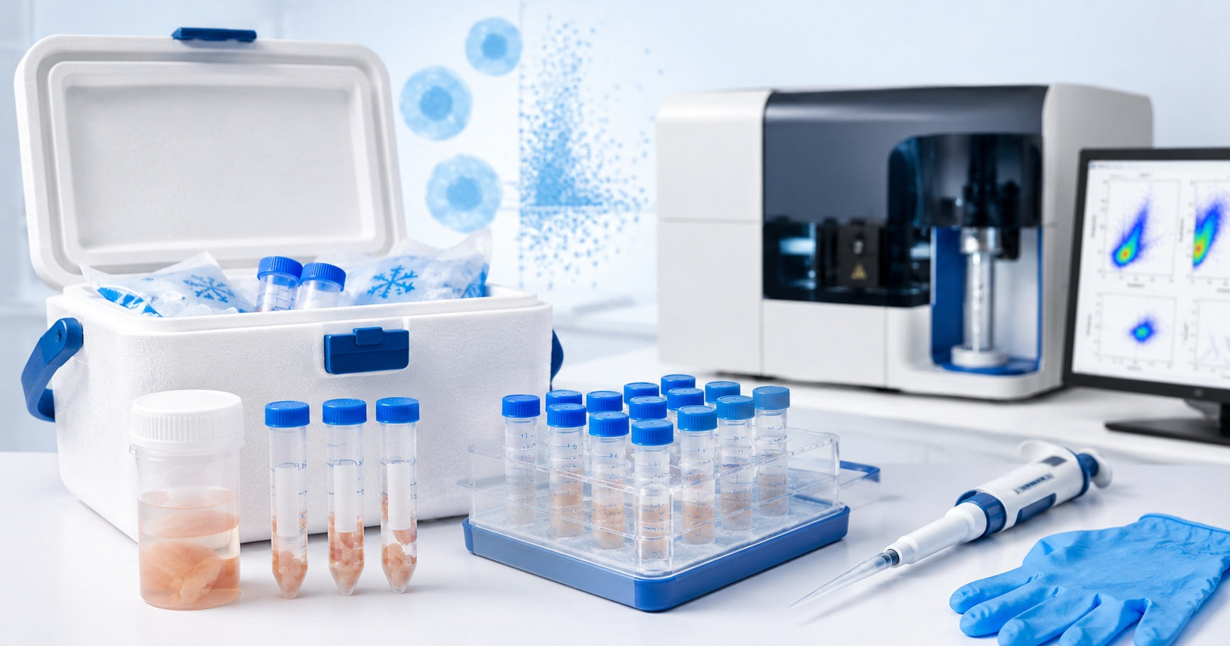
Flow cytometry is a powerful method for analyzing individual cells based on size, granularity, viability, and marker expression. It is widely used in immunology, cancer research, tissue biology, cell sorting, and translational research. For blood or suspension cells, the workflow is usually direct. For solid tissues, the process is more delicate because the sample must be transported, stored, dissociated, filtered, and converted into a high-quality single-cell suspension before analysis.
The transport and storage of tissues for flow cytometry can strongly influence cell viability, cell recovery, surface marker detection, and final data quality. A well-handled tissue sample can produce clean populations, reliable staining, and reproducible results. A poorly transported sample may show low viability, high debris, cell clumping, weak staining, or biased recovery of fragile cell types.
Why Tissue Transport Matters in Flow Cytometry
Flow cytometry depends on healthy, intact single cells. Solid tissues contain cells embedded in extracellular matrix, connective tissue, blood, fat, mucus, necrotic regions, or dense structural material. After collection, cells can experience stress, oxygen changes, temperature changes, enzymatic activity, and mechanical pressure.
Good tissue transport helps slow these changes and preserves the sample until processing. The goal is not only to keep the tissue “available.” The goal is to maintain cells in a condition that supports successful digestion, strong viability, clear antibody staining, and accurate flow cytometry analysis.
Flow Cytometry Needs High-Quality Single Cells
For flow cytometry, cells should be well separated, viable, and free from large clumps. If the tissue is damaged during transport, the final suspension may contain more dead cells, debris, sticky DNA from lysed cells, and aggregates. These problems can affect gating, reduce sorting efficiency, and make results harder to interpret.
Transport Time Affects Cell Viability
Shorter transport and faster processing usually support better cell viability. Longer delays may increase stress, apoptosis, and marker changes. Some tissues tolerate short storage better than others, so each tissue type should be validated under the laboratory’s own workflow.
Tissue Storage Conditions Affect Marker Detection
Surface markers can be sensitive to temperature, digestion enzymes, storage delay, and cell stress. This is especially important when studying immune cell subsets, activation markers, rare populations, or fragile cell types.
Key Goals During Tissue Transport and Storage
A good tissue transport plan should support several goals at the same time.
Maintain Cell Viability
The sample should be kept in conditions that reduce stress and help cells remain intact until processing. Maintaining cell viability during tissue transport improves staining quality, gating confidence, and downstream sorting performance.
Preserve Surface Markers
Many flow cytometry panels depend on surface marker expression. Gentle handling, suitable transport media, and optimized digestion help preserve markers that are important for identifying cell populations.
Prevent Tissue Drying
Fresh tissue samples should not dry out. Drying can damage cells and reduce recovery. Tissue should remain moist in an appropriate medium or buffer during transport.
Reduce Cell Stress and Clumping
Mechanical pressure, long delay, poor temperature control, and tissue damage can increase cell stress. Stressed or dead cells may release DNA, which contributes to cell clumping. A clean transport workflow helps reduce this risk.
Keep Tissue Suitable for Digestion
Tissue digestion works best when the sample is fresh, moist, and not damaged by freezing or harsh handling. Good transport makes the later tissue digestion step more predictable.

Best Transport Media for Tissue Samples
The best transport media for tissue samples depends on the tissue type, transport time, downstream assay, and whether live cells are required.
RPMI-Based Transport Media
RPMI is commonly used for fresh tissue samples intended for flow cytometry. It helps keep cells in a cell-compatible environment during short transport. For many immune profiling workflows, RPMI with protein supplements such as serum or BSA may help support cell condition.
PBS or Saline-Based Transport Buffer
PBS or sterile saline may be acceptable for short transport in some workflows, especially when the tissue will be processed quickly. However, these buffers may not support cell condition as well as complete culture medium during longer transport.
Serum or Protein Supplement
A small amount of serum, BSA, or other protein supplement may help reduce cell stress and surface adhesion. This can be useful for delicate samples, immune cells, or longer handling windows.
Specialized Tissue Preservation Solutions
For studies that require more controlled storage, specialized tissue preservation solutions can help reduce metabolic activity and support short-term preservation. FireGene provides research-focused tissue storage and sample preparation solutions designed to support downstream single-cell workflows.
Recommended Temperature for Tissue Transport
Temperature is one of the most important factors in tissue transport.
Cold Transport for Fresh Tissue
Many fresh tissue workflows use cold transport because low temperature can slow cellular metabolism and reduce stress before processing. Cold transport is commonly done with wet ice or cold packs, but the sample should not freeze.
Avoid Freezing Fresh Tissue for Live-Cell Flow Cytometry
Fresh tissue for live-cell flow cytometry should usually not be frozen unless the protocol is specifically designed and validated for cryopreservation. Freezing can damage cells, reduce viability, and change the recovery of sensitive populations.
Room Temperature Transport May Be Protocol-Specific
Some clinical or laboratory protocols transport certain specimens at room temperature within a defined time window. This is why the receiving laboratory’s SOP should always be followed.
Avoid Temperature Fluctuations
Repeated warming and cooling can stress cells. Use an insulated container, proper packaging, and clear transport instructions to keep conditions consistent.
How Long Can Tissue Be Stored Before Flow Cytometry?
There is no single universal storage time for all tissue types. The best processing window depends on tissue source, density, ischemia time, transport media, temperature, and target cell population.
Ideal Processing Window
For live-cell flow cytometry, fresh tissue should generally be processed as soon as possible. Faster processing helps preserve cell viability and marker expression.
Short-Term Tissue Storage
Short-term storage can be useful when samples are collected at different times or transported from another site. During this period, the sample should remain moist, sterile, and under validated temperature conditions.
Delayed Processing Risks
Long delays can increase dead cells, debris, stress responses, bacterial contamination, and marker changes. These changes may affect flow cytometry gates and reduce confidence in the result.
Validate Each Tissue Type
Tumor tissue, lymph node, skin, lung, liver, brain, gastrointestinal tissue, and blood-rich tissue can behave differently. Each tissue type should have a validated transport and processing plan.
Human Tissue Samples: Special Handling Considerations
Human tissue samples often come from surgery, biopsy, or pathology workflows. These samples may be limited, variable, and time-sensitive.
Fresh Surgical Tissue
Fresh surgical tissue should be placed into suitable transport media quickly after collection. The collection time, transport start time, and processing time should be documented.
Tumor Tissue Samples
Tumor samples may contain necrotic regions, blood, stromal cells, immune cells, and cancer cells. Gentle handling is important because different cell populations may respond differently to transport and digestion.
Lymph Node Tissue
Lymph node tissue can be useful for immune profiling because it contains many immune cells. It should be kept moist and processed carefully to preserve cell populations and marker expression.
Fragile or Blood-Rich Tissue
Some tissues may be fragile, fatty, fibrotic, or rich in red blood cells. These samples may need special washing, red blood cell lysis, optimized digestion, or debris removal before staining.
Biosafety and Documentation
Human tissue samples should be handled according to biosafety, ethical, institutional, and regulatory requirements. Clear labeling and documentation help protect both sample integrity and study quality.
Maintaining Cell Viability During Tissue Transport
Maintaining cell viability during tissue transport is one of the main goals of pre-analytical handling.
Use Suitable Media Volume
The tissue should be covered or immersed enough to prevent drying, but not handled so roughly that cells are damaged. Very small biopsies should not be left exposed to air.
Avoid Tissue Compression
Do not crush tissue inside a tight container. Compression can damage cells and reduce recovery.
Minimize Transport Delay
Plan collection and processing schedules ahead of time. If the tissue is coming from another site, define the maximum accepted transport time before the study begins.
Use Sterile Containers
Sterile, leak-proof containers help reduce contamination risk and keep the sample protected during transport.
Record Transport Conditions
Record collection time, transport media, temperature condition, arrival time, and processing time. These details are useful when comparing viability or troubleshooting inconsistent results.
Tissue Digestion After Transport
Tissue digestion is the step that converts solid tissue into a single-cell suspension for flow cytometry.
Why Tissue Digestion Is Needed
Solid tissue cells are connected by extracellular matrix and cell-cell interactions. Digestion enzymes and gentle mechanical dissociation help release individual cells so they can be stained and analyzed.
Mechanical Dissociation
Mechanical dissociation includes mincing, gentle pipetting, trituration, or using a tissue dissociator. The goal is to break tissue into smaller pieces without causing excessive cell damage.
Enzymatic Digestion
Enzymes such as collagenase, DNase, dispase, and other proteases may be used depending on tissue type. Enzyme selection should be optimized because some enzymes can affect surface markers.
Avoid Over-Digestion
Over-digestion can damage cells, reduce viability, and alter surface epitopes. It may also increase debris.
Avoid Under-Digestion
Under-digestion can reduce cell yield and leave large tissue fragments or clumps. This may lower recovery of important populations.
Filter the Cell Suspension
After digestion, filtering through an appropriate cell strainer helps remove clumps and undigested fragments. FireGene tissue dissociation workflows emphasize gentle digestion, controlled mechanical processing, and filtration to support high-quality single-cell suspensions.
Common Problems Caused by Poor Tissue Transport
Poor transport and storage can affect the entire flow cytometry workflow.
Low Cell Viability
Low viability may result from long delay, drying, freezing, poor media choice, mechanical damage, or harsh digestion.
High Debris
Damaged tissue and dead cells increase debris. This can make gating harder and reduce the clarity of cell populations.
Cell Clumping
Dead cells release DNA, which can make cells sticky. Clumping can block strainers, affect counting, and reduce flow cytometer performance.
Weak Antibody Staining
Poor handling or harsh digestion can reduce surface marker signal. This may make cell populations harder to identify.
Inconsistent Data
When transport conditions vary between samples, flow cytometry results may also vary. Standardized handling improves reproducibility.
Flow Cytometry Workflow After Tissue Arrival
A clear post-arrival workflow helps protect sample quality.
Check Sample Condition
Inspect the tissue on arrival. Note color, dryness, visible blood, necrotic areas, leakage, or transport delay.
Wash Before Digestion
Wash tissue gently to remove excess blood, transport media residue, or loose debris when appropriate.
Digest and Dissociate
Use a tissue-specific digestion method. Control temperature, incubation time, enzyme concentration, and mechanical force.
Count Cells and Check Viability
After digestion and filtration, count cells and measure viability before staining. This helps determine whether the sample is ready for flow cytometry.
Stain With a Suitable Panel
Use optimized antibody panels, Fc blocking when needed, and a live/dead viability dye to improve gating quality.
Best Practices for Multicenter Tissue Transport
When samples come from multiple collection sites, consistency is very important.
Standardize Collection Protocols
Use the same collection container, transport media, temperature range, labeling method, and documentation format across sites.
Define Maximum Transport Time
Set an acceptable time window before starting the study. This helps reduce sample-to-sample variation.
Use Temperature Monitoring
For sensitive studies, temperature tracking can help confirm that samples stayed within the validated range.
Set Acceptance Criteria
Define what qualifies as an acceptable sample. Criteria may include transport time, temperature condition, tissue condition, cell viability, and minimum cell recovery.
Quick Tissue Transport Checklist for Flow Cytometry
Step Best Practice
- Collection Place tissue into a sterile container quickly
- Media Use suitable tissue transport media such as RPMI, saline, or validated preservation solution
- Temperature Maintain validated transport temperature and avoid freezing
- Timing Process fresh tissue as soon as possible
- Digestion Use optimized mechanical and enzymatic dissociation
- Viability Check cell viability before staining
- Clumps Filter the suspension before acquisition
- Documentation Record collection, transport, and processing details
FireGene Support for Tissue Transport and Flow Cytometry Workflows
High-quality flow cytometry data begins with strong sample preparation. Tissue transport, storage, digestion, and single-cell suspension quality all work together to support reliable results.
FireGene supports research workflows with sample preservation, tissue dissociation, cell suspension preparation, and single-cell sample preparation solutions. These tools can help researchers improve consistency when working with fresh tissues before downstream applications such as flow cytometry, cell sorting, and single-cell analysis.
FAQs
What is the best transport medium for tissue samples used in flow cytometry?
RPMI-based culture medium is commonly used for fresh tissue transport before flow cytometry. Sterile saline or PBS may be acceptable for short transport in some protocols, while specialized tissue preservation solutions may be useful for controlled short-term storage.
How do you maintain cell viability during tissue transport?
To maintain cell viability, keep tissue moist, use suitable transport media, avoid freezing, reduce transport delay, prevent compression, use sterile containers, and process the sample as soon as possible.
Should tissue samples be transported on ice for flow cytometry?
Many fresh tissue workflows use cold transport with wet ice or cold packs, but the sample should not freeze. Some protocols may use room temperature transport within a defined time window, so always follow the receiving laboratory’s validated SOP.
Can human tissue samples be stored overnight before flow cytometry?
Some tissue samples may tolerate short-term storage, but overnight storage should be validated for the specific tissue type, marker panel, and downstream goal. Cell viability and marker preservation should be checked before analysis.
Why is tissue digestion important before flow cytometry?
Tissue digestion helps release individual cells from solid tissue so they can be stained and analyzed. A good digestion workflow improves cell yield, viability, and suspension quality.
How does poor tissue storage affect flow cytometry results?
Poor tissue storage can reduce cell viability, increase debris, promote clumping, change marker expression, reduce cell recovery, and make gating less reliable.
What causes low cell viability after tissue digestion?
Delayed transport, poor storage media, freezing, harsh mechanical dissociation, over-digestion, high enzyme exposure, or tissue-specific fragility may cause low viability.
Can tissue samples be frozen before flow cytometry?
Fresh tissue intended for live-cell flow cytometry is usually not frozen unless a validated cryopreservation workflow is being used. Freezing can reduce viability and affect cell recovery.
Conclusion
The transport and storage of tissues for flow cytometry play a major role in sample quality and final data reliability. When fresh tissue samples are kept moist, transported in suitable media, protected from freezing, processed within a validated time window, and digested gently, the final single-cell suspension is more likely to show strong viability, clear staining, and consistent flow cytometry results.
A positive and well-planned workflow helps researchers protect valuable tissue samples from collection to analysis. By standardizing transport media, temperature, timing, digestion, filtration, and viability checks, laboratories can improve cell recovery and generate more dependable flow cytometry data.


