
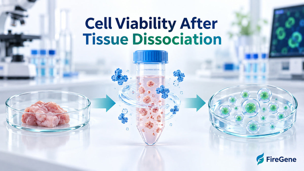
Cell viability after tissue dissociation is measured by identifying intact living cells and calculating their proportion, concentration and total recovery, while an optimized dissociation and cleanup workflow helps create a high-quality cell suspension for downstream research. A successful primary cell isolation workflow balances effective extracellular-matrix digestion with gentle cellular handling. Enzyme selection, incubation time, mechanical force, temperature, washing, debris removal and counting consistency can all influence the final result.
Researchers can use the FireGene Multi Tissue Dissociation Kit to generate single-cell suspensions from diverse mammalian tissues. When additional sample refinement is beneficial, FireGene solutions for dead-cell removal, debris cleanup and aggregation control can help prepare cells for flow cytometry, tissue culture, primary cell culture and single-cell sequencing.
FireGene Solutions for Dissociation and Cell-Suspension Quality
|
Workflow requirement |
FireGene product |
Application |
|
Multi-tissue dissociation |
Enzymatically converts diverse mammalian tissues into single-cell suspensions |
|
|
Viable-cell enrichment |
Supports enrichment of viable cells from tissue-derived suspensions |
|
|
Dead-cell and debris cleanup |
Helps prepare cleaner samples for counting and downstream analysis |
|
|
Aggregation management |
Supports a uniform, well-dispersed cell suspension |
The best product combination depends on the tissue type, starting condition, cell population and downstream application.
Why Cell Viability Matters After Tissue Dissociation
Tissue dissociation releases individual cells from a structured extracellular environment. The quality of the resulting suspension can influence every subsequent stage, including cell counting, fluorescence staining, cell sorting, culture and sequencing.
A high-quality suspension generally contains:
- Intact cells with preserved membranes
- A useful concentration of viable cells
- Limited tissue debris
- Well-dispersed individual cells
- Minimal large aggregates
- Representative recovery of the desired populations
Cell viability provides an important quality-control measurement, but it becomes more informative when interpreted with viable-cell concentration and total recovery.
Cell Viability Percentage
Cell viability percentage describes the proportion of counted cells classified as viable:
Cell viability (%) = Number of viable cells ÷ Total number of cells × 100
For example, a sample containing 800 viable cells among 1,000 total counted cells has 80% viability.
This metric offers a convenient overview of membrane integrity within the measured suspension. It is especially useful for comparing different incubation times, tissue dissociation methods and sample-handling conditions.
Viable-Cell Concentration
Viable-cell concentration indicates how many living cells are present per unit of volume. This information is essential when a downstream assay requires a defined cellular input. A sample can have an excellent viability percentage while containing a modest total number of cells. Recording viable-cell concentration provides the additional information required for loading, plating and normalization.
Total Viable-Cell Recovery
Total viable-cell recovery measures the total number of living cells obtained from the tissue:
Total viable-cell yield = Viable-cell concentration × Final suspension volume
When an estimate of the original target-cell number is available, researchers can also calculate recovery:
Viable-cell recovery (%) = Final viable-cell number ÷ Estimated starting viable-cell number × 100
Competitor guides frequently emphasize viability percentage alone. Recording viability, concentration and total viable yield gives researchers a more complete view of dissociation performance.
How to Measure Cell Viability After Tissue Dissociation
Several cell counting and viability measurement methods are available. Method selection depends on sample complexity, available instruments, required throughput and the desired level of cellular specificity.
Trypan Blue Dye-Exclusion Method
Trypan Blue staining evaluates membrane integrity. Viable cells with intact membranes exclude the dye and remain relatively clear, while cells with permeable membranes absorb it and appear blue.
A typical workflow includes:
- Gently mix the cell suspension.
- Combine the sample with Trypan Blue at a standardized ratio.
- Load the mixture into a hemocytometer or automated counting slide.
- Count clear and blue-stained cells.
- Apply the relevant dilution factor.
- Calculate viability and viable-cell concentration.
Trypan Blue viability assessment is familiar, accessible and suitable for many routine cell preparations. Consistent stain ratio, incubation interval and counting time support comparable results across samples.
Manual Cell Counting with a Hemocytometer
A hemocytometer contains a defined grid and chamber depth, allowing cell concentration to be calculated from a known volume. During manual cell counting, researchers count viable and dye-positive cells in selected grid areas. Counts from multiple squares can be averaged before applying chamber volume and dilution factors.
For a standard Neubauer chamber, cell concentration is commonly calculated as:
Cells/mL = Average cells per large square × Dilution factor × 10⁴
A representative suspension is important because tissue-derived samples may settle or develop local differences in cell density. Gentle mixing immediately before chamber loading helps distribute cells evenly. Counting technical replicates also supports a clearer assessment of sample uniformity.
Automated Brightfield Cell Counting
Automated brightfield counters capture images and classify objects according to size, shape, contrast and staining intensity. These systems can rapidly report:
- Total-cell concentration
- Viable-cell concentration
- Dead-cell concentration
- Viability percentage
- Cell-size distribution
Brightfield counting can increase throughput and reduce operator-dependent variation. Appropriate size gates and detection settings help the instrument distinguish cells from tissue fragments, apoptotic bodies and other particles. Researchers working with complex samples can inspect captured images rather than relying only on the final percentage.
Fluorescence-Based Viability Measurement
Fluorescence-based counting uses dyes that distinguish nucleated cells and cells with permeable membranes. AO/PI staining is one widely used approach: acridine orange identifies nucleated cells, while propidium iodide identifies membrane-compromised cells.
This method can offer added cellular specificity in samples containing:
- Tissue debris
- Red blood cells
- Irregularly shaped primary cells
- Small particles
- Mixed cellular populations
For a detailed method comparison, internally link this section to your published Fluorescence vs Trypan Blue Automated Cell Counting guide.
Flow Cytometry-Based Viability Assessment
Flow cytometry can evaluate individual cells using fluorescent viability dyes alongside surface or intracellular markers. This approach is valuable when viability must be measured within a particular cell population rather than across the complete suspension. For example, a heterogeneous tissue suspension may have strong overall viability while one specific population shows a different recovery profile. Population-level analysis can reveal this valuable distinction.

Factors That Influence Cell Viability During Tissue Dissociation
Cell viability reflects the complete preparation workflow. Standardizing the following variables helps researchers generate reproducible suspensions.
Tissue Collection and Processing Time
Prompt tissue processing supports cellular integrity. Researchers can define a consistent interval between tissue collection, transport and dissociation. When immediate processing is unavailable, a validated preservation approach can help maintain sample quality. Time, temperature and transport medium should be recorded for each sample.
Tissue Size and Mechanical Preparation
Cutting tissue into small, consistent pieces increases the surface area available for enzymatic digestion. Uniform fragments promote even processing and make it easier to compare samples. Gentle mechanical support can then help release cells as the extracellular matrix softens. Controlled pipetting, shaking or instrument-based processing supports effective dissociation while preserving the desired cell populations.
Enzyme Selection
Collagenase and other proteolytic enzymes are widely used in tissue dissociation because extracellular matrix composition varies between tissues. The ideal enzyme system depends on matrix density, tissue age, fibrosis, target-cell sensitivity and the surface markers required downstream. The FireGene Multi Tissue Dissociation Kit uses an optimized enzyme blend and buffer system for diverse mammalian tissues. Its listed compatible samples include liver, lung, kidney, intestine, tumor, ovary, skin, lymphoid and embryonic tissues. For specialized tissues with extensive calcification or high lipid content, a tissue-specific method may offer the most suitable processing route.
Digestion Time and Temperature
Sufficient digestion promotes efficient cell release, while timely monitoring helps preserve cellular condition. Rather than selecting one universal incubation period, researchers can observe both tissue-clump reduction and cell viability during processing. The FireGene product guidance recommends beginning quality checks after 15 minutes and repeating them every 20–30 minutes when appropriate. This allows digestion time to be matched to the tissue’s actual response. Maintaining the recommended temperature and mixing pattern also supports enzyme consistency.
Pipetting and Centrifugation
Controlled pipetting assists cell release and suspension uniformity. Smooth, reproducible pipetting is especially valuable for primary cells and sensitive tissue-derived populations. Centrifugation conditions can be matched to cell size and density. Using a validated relative centrifugal force, duration and acceleration setting supports efficient collection while maintaining cell quality.
Residual Enzymes and Washing
Once the desired level of dissociation is achieved, washing helps remove residual enzyme solution, soluble debris and other process materials. The FireGene Multi Tissue Dissociation Kit instructions include filtration and defined washing steps. Its product guidance explains that complete washing supports cleaner counting and helps limit continued enzyme exposure. Using the recommended wash medium also provides a consistent transition from digestion to counting or downstream preparation.
Cell Aggregation
Extracellular DNA released by membrane-compromised cells can contribute to cell aggregation. Cell clusters may affect counting accuracy, filtration, antibody staining and microfluidic loading. Gentle mixing and appropriate suspension conditions encourage a uniform sample. The FireGene Anti-Clumping Agent can also support aggregation management when compatible with the selected cell type and assay.

How to Improve Cell Viability After Tissue Dissociation
An effective optimization plan evaluates the complete path from intact tissue to the final assay.
Match the Method to the Tissue
Tissues differ in extracellular matrix density, cell composition and sensitivity. A liver, brain, tumor or fibrotic specimen may each benefit from different tissue dimensions, incubation intervals and mechanical processing. Begin with the recommended protocol and adjust one variable at a time. This approach makes improvements easier to identify and reproduce.
Monitor Dissociation Progress
Periodic visual examination helps researchers identify when tissue fragments have reduced sufficiently and individual cells are being released.
A small aliquot can be evaluated for:
- Remaining tissue clumps
- Cell dispersion
- Membrane integrity
- Visible debris
- Aggregate formation
- Viable-cell concentration
This real-time approach creates a more informative endpoint than relying on incubation time alone.
Use Gentle and Consistent Handling
Consistent handling improves experimental comparability. Predefine the tissue-cutting method, pipette size, number of mixing cycles, filtration step and centrifugation program. Keeping these variables stable helps distinguish tissue-related biological variation from process-related variation.
Remove Dead Cells When Beneficial
A sample may contain enough viable cells for the next step while also carrying dead cells that influence counting or downstream analysis. The FireGene Dead Cell Removal Solution supports density-based viable-cell enrichment from mammalian tissue-derived suspensions. Researchers can perform an initial count, complete cleanup and then repeat the count to quantify final viable-cell recovery. This before-and-after measurement demonstrates the practical value of the cleanup step.
Remove Dead Cells and Tissue Debris Together
Some dissociated tissues contain both nonviable cells and extracellular fragments. The FireGene Dead Cell Debris Removal Kit supports removal of both components while enriching intact cells. Cleaner suspensions can support clearer microscopic images, more consistent cell classification and efficient preparation for downstream workflows.
Maintain a Uniform Single-Cell Suspension
After cleanup, resuspend the cell pellet evenly and inspect the sample for aggregates. A uniform suspension supports representative aliquoting, accurate cell counting and consistent assay input. When cell settling occurs, mix gently before every replicate and counting-chamber load.
Measure Cells Promptly
Cells continue responding to their environment after dissociation. A consistent interval between final resuspension, staining and cell counting helps make results comparable. When a short delay is required, use validated temperature, medium and concentration conditions. Repeat the viability measurement before downstream use when the storage interval may have influenced the sample.

A Practical Dissociation-to-Viability Workflow
- Define the downstream application: Establish cell number, viability, purity and concentration requirements.
- Select a tissue-appropriate method: Match enzymatic and mechanical processing to the sample.
- Prepare uniform tissue pieces: Use consistent dimensions to support even digestion.
- Begin enzymatic dissociation: Maintain the specified temperature and mixing conditions.
- Monitor the sample: Examine cell release, tissue clumps and viability at defined intervals.
- Stop digestion at the appropriate endpoint: Transition promptly into filtration and washing.
- Filter the suspension: Remove remaining tissue fragments with a suitable cell strainer.
- Complete the required washes: Remove residual dissociation reagents and soluble material.
- Perform the initial count: Record total cells, viable cells, viability and concentration.
- Apply cleanup when useful: Remove dead cells, debris or aggregates according to sample condition.
- Repeat viability measurement: Calculate final viable-cell yield and recovery.
- Normalize downstream input: Adjust the sample to the required viable-cell concentration.
This workflow connects tissue processing, cell viability measurement and product selection in one quality-control strategy.

Cell Viability Troubleshooting Guide
|
Observation |
Possible workflow factor |
Practical optimization |
|
Strong viability with modest cell yield |
Partial tissue digestion or cell loss during washing |
Review tissue-piece size, digestion endpoint and transfer recovery |
|
Useful cell yield with visible debris |
Remaining extracellular material |
Add an appropriate debris-cleanup step |
|
Variable replicate counts |
Settling or uneven suspension |
Mix gently before each measurement and load chambers consistently |
|
Cell aggregates |
Released DNA or incomplete dispersion |
Optimize filtration, mixing and anti-clumping support |
|
Inconsistent dye-exclusion results |
Variable staining interval |
Standardize dye ratio and measurement timing |
|
Many remaining tissue clumps |
Digestion needs further optimization |
Monitor digestion progressively while tracking cell viability |
|
Downstream loading variation |
Concentration calculated from total cells alone |
Normalize using viable-cell concentration |
This table adds a practical decision layer that many general tissue dissociation guides do not provide.
Frequently Asked Questions
What is a good cell viability percentage after tissue dissociation?
A suitable viability percentage depends on the tissue, cell type and downstream assay. Researchers should compare the result with their application’s validated acceptance criteria. Viable-cell concentration and total viable yield should be evaluated alongside the percentage.
How soon should cells be counted after dissociation?
Cells are best counted at a consistent, validated time after final washing and resuspension. Prompt counting creates a reliable picture of the prepared sample and supports accurate downstream normalization.
How does collagenase support primary cell isolation?
Collagenase helps digest collagen-rich extracellular matrices and release individual cells. Enzyme type, concentration, temperature and incubation time can be optimized for the tissue and target population.
Can tissue debris affect cell counting?
Yes. Tissue fragments and apoptotic particles may be detected as cell-like objects, particularly during brightfield counting. Image review, suitable instrument settings and sample cleanup support accurate classification.
Is Trypan Blue suitable for tissue-derived primary cells?
Trypan Blue can be used for many primary-cell suspensions. Optimized staining time, size gates, representative sampling and careful handling help produce consistent measurements.
What is the difference between viability and recovery?
Viability is the percentage of counted cells that are alive. Recovery describes how many viable cells remain after dissociation or cleanup relative to the starting estimate. Together, they show both sample condition and usable yield.
Should dead cells be removed before single-cell sequencing?
Dead-cell removal can be valuable when the starting suspension contains a meaningful nonviable fraction. Researchers should measure viable-cell recovery after cleanup and confirm that the final sample meets the platform’s requirements.
Should viability be measured before and after cleanup?
Yes. A pre-cleanup count characterizes the starting suspension, while a post-cleanup count shows changes in viability, concentration and viable-cell recovery. This comparison supports data-driven workflow optimization.
Conclusion
Cell viability after tissue dissociation is best evaluated through a combination of viability percentage, viable-cell concentration and total viable-cell recovery. Gentle enzymatic dissociation, progressive quality checks, complete washing, representative counting and targeted sample cleanup can create a strong foundation for reliable downstream research.
FireGene supports this workflow with solutions for multi-tissue dissociation, viable-cell enrichment, dead-cell and debris removal, and aggregation management. Start with the FireGene Multi Tissue Dissociation Kit, browse the Cell Cleanup and Suspension Optimization collection, or contact FireGene to discuss a tissue-specific sample-preparation workflow.


