
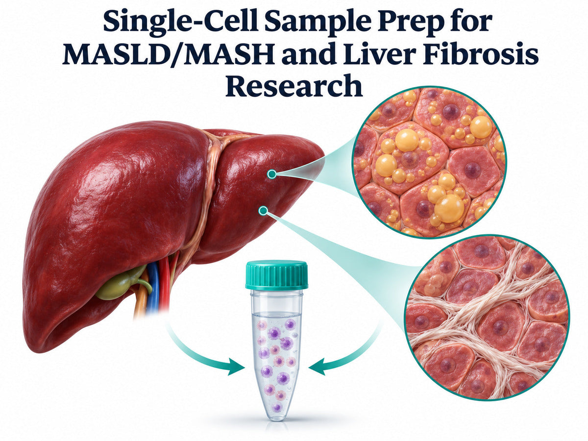
Quick answer: For MASLD/MASH (formerly NAFLD/NASH) and liver fibrosis single-cell studies, choose your preparation around the specimen and the populations you need to recover. Use scRNA-seq when fresh tissue yields a suitable intact-cell suspension. Consider snRNA-seq for frozen liver or when fragile, lipid-laden hepatocytes are underrepresented. Pilot the workflow before processing the full cohort.
FireGene Liver Sample Preparation Kit Catalog (FG-BA3323 and FG-BA3339)
|
Product |
Catalog number |
When to choose it |
Pack formats listed |
Order |
|
Liver Dissociation Kit |
FG-BA3323 |
Prepare an intact-cell suspension from fresh liver tissue |
10 or 50 reactions |
|
|
Dead Cell Debris & Impurities Removal Kit |
FG-BA3339 |
Clean up an existing cell suspension when dead cells or debris interfere with sample quality |
2-reaction option shown. Components are also documented for 10- and 50-reaction formats. |
Why Choose FireGene for Liver Single-Cell Sample Prep?
Start with FG-BA3323 to prepare a liver cell suspension. Evaluate FG-BA3339 when your pilot identifies a need for cleanup. Compare viable yield and target-cell recovery before and after cleanup to see whether it improves the sample you plan to load. The certificate for lot FG250821 records a pass against release criteria of at least 85% viable hepatocytes after digestion and at least 1 million viable cells per 100 mg of mouse liver. These thresholds describe lot acceptance. Measure yield, viability, and cell-type representation in the fatty or fibrotic specimens you plan to study. Liver Dissociation Kit and lot documentation
Why Does Single-Cell Profiling Matter in MASH and Liver Fibrosis?
Liver fibrosis single-cell RNA sequencing helps identify which populations express disease-associated genes. An increase in collagen-related expression, for example, can reflect a shift in the abundance of matrix-producing cells, a change in expression within those cells, or both. That distinction matters when you compare healthy, steatotic, and fibrotic specimens. Human liver atlases describe scar-associated macrophages, activated hepatic stellate cells, portal fibroblasts, and disease-associated endothelial populations. Mouse fibrosis studies also describe communication between stellate cells and macrophages. If your study focuses on that interaction, check that your pilot recovers enough cells from both populations to support the comparison.
Which Mouse Models Present Different Sample Preparation Challenges?
Pilot the workflow on specimens that represent the groups you plan to compare. A healthy control establishes a handling baseline. A specimen from the disease group shows how the preparation performs at the degree of steatosis or fibrosis relevant to the experiment. Where possible, link sequencing samples to matched histology.
|
Mouse model |
What it helps you study |
Sample preparation priority |
|
GAN diet in an obese mouse background |
Steatosis and fibrosis in a model with metabolic dysfunction |
Check recovery of large, lipid-laden hepatocytes. Compare intact cells and nuclei if hepatocyte states are central to the study. |
|
CDAHFD |
Fibrosis in a choline-deficient, amino acid-defined high-fat diet model |
Include representative fibrotic tissue in the pilot. Assess residual tissue, viable recovery, and representation of stellate and immune populations. |
|
Western diet plus CCl₄ |
Steatohepatitis and fibrosis in a combined diet and injury model |
Record the tissue region and compare cell recovery across groups before interpreting changes in inflammatory or stromal populations. |
When comparing models or interpreting a human MASH liver biopsy dataset alongside mouse data, record the specimen type, disease model, and preparation method. Differences in these factors can affect which cells appear in the dataset.

How Should You Adjust Dissociation for Fatty and Fibrotic Liver?
Fatty liver. Steatotic hepatocytes can be large and fragile, making them harder to recover during dissociation or capture on some platforms. Track hepatocyte recovery alongside overall viability. If you see viable hepatocytes before loading but recover few in the dataset, investigate capture as well as tissue preparation. A matched nuclei pilot can help you assess hepatocyte representation.
Fibrotic liver. Assess residual tissue and usable cell recovery together. Compare preparations with similar tissue inputs and record how each sample was handled. If tissue fragments persist, review the kit instructions and evaluate the preparation on a small set of representative specimens. Select the approach that recovers the cell populations you need and meets your sequencing core’s intake criteria.
Cleanup. Compare matched aliquots before and after dead-cell and debris removal. Record the percentage of viable cells and the absolute number of viable cells. If you study a scarce population, check its recovery as well. An increase in the viability percentage can coincide with a decrease in the number of usable target cells.

scRNA-seq vs. snRNA-seq for Fibrotic Liver
Choose between single-cell RNA sequencing and single-nucleus RNA sequencing based on your specimen and the readout you need. A matched human liver study used fresh tissue dissociates for scRNA-seq and snap-frozen tissue for snRNA-seq. The two approaches provided complementary views of liver cell populations and gene expression.
|
Study situation |
Approach to evaluate first |
What to check in the pilot |
|
Fresh tissue yields viable cells, and the study also needs surface phenotyping |
scRNA-seq with a compatible surface phenotyping workflow |
Viability, aggregates, and target-cell representation after capture |
|
Archived snap-frozen liver is the available material |
snRNA-seq |
Nuclei integrity, debris, and recovery across specimens |
|
Steatotic hepatocytes are central to the study and underrepresented in the intact-cell pilot |
Matched cells-versus-nuclei pilot |
Hepatocyte recovery and cell-state representation in each dataset |
|
The study targets rare stellate or scar-associated immune populations |
Broad profiling plus a targeted pilot, where tissue permits |
Whether enrichment recovers enough additional target cells for the analysis |
Use the same preparation across comparison groups where possible. If you combine scRNA-seq and snRNA-seq data, record the modality for each specimen and account for it during analysis. Where your design permits, include matched specimens so you can assess how modality affects the populations you recover.

What Should You Check Before Loading Samples?
Ask your sequencing core for the acceptance criteria for your platform and assay. For intact-cell suspensions, record viability, concentration, viable yield, aggregates, and debris. For nuclei, record concentration, integrity, clumps, and visible contamination. Use the same counting method across comparable specimens. After pilot sequencing, review cell-type recovery by specimen. Check whether hepatocytes, hepatic stellate cells, Kupffer cells or other macrophages, and liver sinusoidal endothelial cells are represented at the level your question requires. For studies of stress or activation, document processing time and temperature. Research in other tissues identified dissociation-associated changes in FOS and JUN, making handling records relevant when interpreting these signals. How Should You Interpret Fibrosis Markers and Spatial Context?
Assess fibrosis-related expression within annotated cell populations. COL1A1, COL1A2, and COL3A1 can help characterize matrix-producing states. TREM2 and CD9 have been reported in scar-associated macrophages in human cirrhosis. Interpret these markers in the context of the species, disease model, and broader expression profile. If your question concerns where these populations localize, pair the transcriptomic results with histology or spatial data. A liver single-cell dataset shows which populations you captured and what they express. Tissue-based measurements help determine where those populations occur relative to scar regions.

Frequently Asked Questions
Can you use frozen liver tissue for single-cell studies?
Yes. snRNA-seq can profile nuclei isolated from frozen liver. Confirm that the storage conditions and nuclei preparation meet your sequencing core’s requirements. Use a pilot to check nuclei integrity and representation of your target populations before processing the rest of the cohort.
Why are hepatocytes underrepresented in some MASH scRNA-seq datasets?
Large, fragile, lipid-laden hepatocytes can be lost during dissociation or underrepresented during capture. Compare the hepatocytes observed before loading with those identified after sequencing. If intact-cell recovery is insufficient for your study, evaluate a matched nuclei preparation.
Should you enrich hepatic stellate cells for fibrosis research?
Consider enrichment when stellate cell states are the main endpoint and a mixed pilot recovers too few cells for the planned comparison. Keep a matched mixed aliquot if the study also examines relationships between stellate cells, macrophages, and other liver populations.
What should you include in a kit quote request?
Include the species, model, tissue format, planned number of preparations, and target populations. State whether you plan to profile intact cells or nuclei and whether your pilot showed a need for debris removal. Ask for pack availability and the applicable lot documentation. Request a quote
References
- Resolving the fibrotic niche of human liver cirrhosis at single-cell level. Nature, 2019.
- Single-cell RNA sequencing reveals the heterogeneity and intercellular communication of hepatic stellate cells and macrophages during liver fibrosis. MedComm, 2023.
- Protocol for minimized-bias profiling of liver and visceral adipose tissue in mice using integrated single-nucleus RNA sequencing. STAR Protocols. PMCID PMC13356638.
- Single-Cell, Single-Nucleus, and Spatial RNA Sequencing of the Human Liver Identifies Cholangiocyte and Mesenchymal Heterogeneity. Hepatology Communications, 2022.
- A Simple Diet- and Chemical-Induced Murine NASH Model with Rapid Progression of Steatohepatitis, Fibrosis and Liver Cancer. PMID 29572095.
- An improved mouse model that rapidly develops fibrosis in non-alcoholic steatohepatitis. PMID 23305254.
- A human single-cell atlas identifies OLR1+ scar-associated macrophages as a potential therapeutic target for chronic liver disease. Nature Genetics, 2026.
- Metabolic and epigenetic abnormalities cause hepatic fibrogenesis in metabolic dysfunction–associated steatohepatitis model mice. Journal of Biological Chemistry.
- Dissociation of solid tumor tissues with cold active protease for single-cell RNA-seq minimizes conserved collagenase-associated stress responses. Genome Biology, 2019.
Order Reagents for Your Liver Fibrosis Single-Cell Study
Choose FG-BA3323 for your intact-cell preparation pilot. Add FG-BA3339 if dead cells or debris warrant a cleanup step. Include your tissue format, sample count, and target populations when requesting a quote.
Shop Liver Dissociation Kit · Shop Dead Cell Debris Removal Kit · Browse Sample Preparation Kits · Request a Quote


