
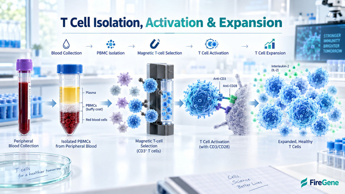
T cells are commonly isolated by preparing PBMCs from peripheral blood or leukapheresis material, enriching the required T-cell population, activating the selected cells with receptor-directed signals and expanding them in a controlled ex vivo culture. A productive T-cell workflow begins with clean, viable starting material. The quality of blood collection, PBMC isolation, cell separation and suspension preparation can influence cell recovery, activation consistency and expansion performance.
The FireGene Human PBMC Isolation Solution supports the preparation of peripheral blood mononuclear cells before T-cell selection. Complementary FireGene solutions can help researchers manage residual red blood cells, dead cells, debris and cellular aggregation before subsequent research steps.
FireGene Products for Pre-Selection Sample Preparation
|
Workflow need |
FireGene product |
Research value |
|
Preparing PBMCs |
Provides a mononuclear-cell starting population before T-cell selection |
|
|
Managing residual red blood cells |
Supports RBC removal when required |
|
|
Enriching viable cells |
Helps prepare a viable cellular suspension |
|
|
Removing dead cells and debris |
Supports cleaner samples for downstream processing |
|
|
Maintaining uniform suspensions |
Helps manage cellular aggregation |
These FireGene products support PBMC and sample preparation before T-cell selection. Selection beads, activation reagents and expansion conditions should be sourced and validated separately for the intended research application.
T-Cell Isolation and Expansion: Quick Overview
|
Workflow stage |
Main objective |
Common approach |
|
Starting-material preparation |
Obtain mononuclear cells |
PBMC isolation from peripheral blood or leukapheresis material |
|
T-cell selection |
Enrich the required population |
Positive or negative selection |
|
Quality assessment |
Measure sample condition |
Cell counting, viability and purity analysis |
|
T-cell activation |
Deliver activating signals |
Antibody-coated beads or antigen-presenting cells |
|
Ex vivo expansion |
Increase viable cell numbers |
Controlled culture with appropriate cytokine support |
|
Final assessment |
Confirm cellular quality |
Viability, phenotype, purity and functional testing |
What Are T Cells?
T cells are lymphocytes that support adaptive immune responses. They recognize specific signals, coordinate immune activity and contribute to the removal of selected target cells.
Major T-Cell Subsets
Common T-cell subsets include:
- CD4+ helper T cells: Coordinate immune responses through signaling and cytokine production.
- CD8+ cytotoxic T cells: Recognize and respond to selected target cells.
- Regulatory T cells: Support balanced immune activity.
- Naive T cells: Provide a starting population capable of responding to new antigens.
- Memory T cells: Support rapid responses following previous antigen exposure.
Researchers isolate these populations for immune profiling, functional assays, disease modeling, drug discovery and cell-therapy research.
Where Are T Cells Obtained?
Peripheral Blood
Peripheral blood contains red blood cells, platelets, granulocytes and peripheral blood mononuclear cells. A PBMC preparation concentrates lymphocytes and monocytes into a practical starting fraction for immune cell isolation.
Leukapheresis Material
Leukapheresis selectively collects leukocytes from circulating blood while returning other components to the donor. It can provide a larger starting population than a routine blood draw, making it valuable when substantial cell numbers are required. Blood collection and leukapheresis are distinct collection approaches, but both can supply starting material for T-cell research.
How Are PBMCs Prepared Before T-Cell Isolation?
Density-based centrifugation is commonly used to separate peripheral blood mononuclear cells from other blood components. After centrifugation, the PBMC layer is collected, washed and resuspended for counting and further cell separation. The FireGene Human PBMC Isolation Solution is designed to prepare lymphocyte and PBMC suspensions from diluted human peripheral blood. The resulting cells can support research applications including immune profiling, flow cytometry, cell culture and single-cell sequencing.
Assessing the Starting Suspension
Before T-cell selection, researchers can record:
- Total-cell concentration
- Viable-cell concentration
- Cell viability
- Visible debris
- Cellular aggregation
- Starting sample volume
These measurements establish a baseline for calculating recovery after cell separation.

How Are T Cells Isolated?
T cell isolation enriches the required population from PBMCs or another leukocyte-containing sample. Magnetic-activated cell separation and fluorescence-activated cell sorting are two common approaches.
Positive T-Cell Selection
Positive selection directly captures T cells expressing a chosen surface marker. Antibody-coated beads may recognize markers such as CD3, CD4 or CD8. The labeled target cells respond to a magnetic field and are separated from the remaining population. This method provides direct enrichment when a clear target marker is available.
Negative T-Cell Selection
Negative selection labels and removes unwanted leukocytes while leaving the target T cells comparatively untouched. This approach can be useful when researchers want target-cell receptors to remain available for subsequent staining or functional analysis. For a detailed comparison, internally link this section to your article on positive vs negative selection for immune cell isolation.
Fluorescence-Activated Cell Sorting
Fluorescence-activated cell sorting identifies cells using multiple fluorescent markers and separates defined populations at the individual-cell level. It supports precise isolation of T-cell subsets with specific phenotypic profiles. The preferred T cell isolation method depends on required purity, recovery, available markers, processing time and downstream use.

How Are T Cells Activated?
T-cell activation occurs when cells receive receptor-directed and co-stimulatory signals. This stage prepares selected T cells for proliferation and functional activity.
Antibody-Coated Activation Beads
T-cell activation beads commonly present antibodies directed toward molecules such as CD3 and CD28. These provide receptor and co-stimulatory signals in a standardized research format.
Researchers can optimize:
- Bead-to-cell ratio
- Starting viable-cell density
- Activation duration
- Culture medium
- Mixing conditions
- T-cell subset
- Downstream objective
Antigen-Presenting Cells
Antigen-presenting cells can stimulate T cells by presenting specific antigens together with appropriate co-stimulatory signals. This approach can be valuable when antigen-specific T-cell responses are central to the research question. Activation success can be evaluated through cell-surface markers, cytokine production, proliferation and functional response.

How Are T Cells Expanded Ex Vivo?
T cell expansion increases the number of viable cells under controlled culture conditions. After activation, cells are transferred into an appropriate culture system with medium, nutrients and cytokine support matched to the desired population.
Control the Culture Environment
Researchers can standardize:
- Temperature and gas conditions
- Culture vessel
- Initial viable-cell concentration
- Feeding schedule
- Cytokine combination
- Culture duration
- Sampling intervals
Maintaining an appropriate viable cell density supports consistent access to nutrients and activation signals.
Monitor T-Cell Expansion
Useful expansion measurements include:
- Viable-cell concentration
- Cell viability
- Total viable-cell number
- T-cell purity
- Subset composition
- Activation phenotype
- Functional activity
Expansion can be reported using:
Fold expansion = Final viable-cell number ÷ Starting viable-cell number
For example, increasing from one million viable T cells to ten million represents a tenfold expansion. This calculation provides more useful information than reporting final concentration alone because it connects the result with the actual starting population.

Why Sample Quality Matters Before T-Cell Selection
Dead cells, residual red blood cells, debris and cellular aggregates can affect counting, antibody labeling and sample handling. A clean, uniform starting suspension supports efficient contact between cells and selection reagents.
A Practical T-Cell Isolation and Expansion Workflow
- Collect the starting material: Obtain peripheral blood or leukapheresis material.
- Prepare PBMCs: Separate the mononuclear-cell fraction through a validated centrifugation workflow.
- Assess sample quality: Measure concentration, viability, debris and aggregation.
- Apply cleanup when beneficial: Manage red blood cells, dead cells or debris.
- Select the T-cell population: Choose positive selection, negative selection or fluorescence-based sorting.
- Confirm selection performance: Measure purity, viable-cell recovery and concentration.
- Activate the cells: Apply a validated receptor-directed activation method.
- Begin ex vivo expansion: Culture the cells under suitable conditions.
- Monitor the culture: Record viability, density, phenotype and fold expansion.
- Evaluate final cells: Confirm that the population meets the research workflow’s requirements.
Research Workflows and Clinical Manufacturing
Research-scale T cell production supports laboratory studies, assay development and process optimization. Clinical manufacturing follows additional requirements involving qualified materials, controlled facilities, process validation, traceability, sterility testing and defined release criteria. CAR T-cell production also includes specialized stages such as genetic modification and extensive quality control. Research-use sample-preparation products should therefore be presented separately from materials qualified for clinical manufacturing.
Frequently Asked Questions
How are T cells isolated from blood?
Blood is commonly processed to prepare PBMCs, after which the required T cells are enriched through magnetic selection or fluorescence-activated cell sorting.
What is the difference between T-cell isolation and activation?
Isolation separates the desired T cells from other cells. Activation provides the signals that prepare selected cells for proliferation and functional activity.
Are T cells isolated directly from whole blood?
Some systems can process whole blood, while many workflows first prepare PBMCs to create a concentrated mononuclear-cell starting population.
How does magnetic T-cell selection work?
Antibody-coated magnetic particles label either the desired T cells or unwanted populations. A magnetic field then separates labeled and unlabeled cells.
How is T-cell expansion measured?
Researchers can monitor viable-cell concentration, total viable-cell number, viability, phenotype, and fold expansion throughout the culture period.
Can leukapheresis material be used for T-cell isolation?
Yes. Leukapheresis material provides a leukocyte-rich starting sample that can be processed for T-cell selection and research-scale expansion.
Conclusion
T-cell isolation and expansion is a connected process that begins with blood-derived material and progresses through PBMC preparation, T-cell selection, activation, controlled culture and final quality assessment. A clean and viable starting suspension supports every subsequent stage. FireGene provides research solutions for PBMC isolation, red blood cell management, dead-cell and debris removal, and aggregation control before T-cell selection. Explore FireGene PBMC isolation products or contact FireGene to discuss a sample-preparation workflow for your T-cell research.


