
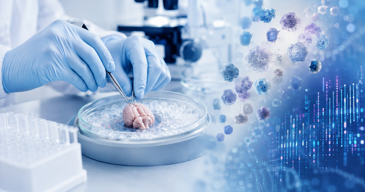
Dissociation of microdissected mouse brain tissue for single-cell RNA sequencing requires a gentle, standardized workflow that supports viable brain-cell recovery, representative gene-expression profiles and a clean suspension for downstream scRNA-seq. Precise microdissection, controlled enzymatic digestion, careful mechanical handling, efficient debris management and accurate cell counting can improve sample consistency for advanced neuroscience research.
Mouse brain tissue contains diverse populations, including neurons, astrocytes, oligodendrocytes, endothelial cells and microglia. Its complex structure and abundant myelin make a brain-specific preparation workflow especially valuable. Optimized tissue dissociation helps researchers obtain a uniform cell suspension suitable for single-cell RNA sequencing, flow cytometry, cell sorting and other cell biology applications.
How Is Microdissected Mouse Brain Tissue Prepared for scRNA-seq?
A well-organized mouse brain tissue dissociation protocol generally includes:
- Collecting and preserving the brain tissue
- Microdissecting the required anatomical region
- Mincing the tissue into consistent fragments
- Applying controlled enzymatic digestion
- Performing gentle mechanical trituration
- Filtering the resulting cell suspension
- Separating myelin and tissue debris
- Completing optional viability-based cleanup
- Measuring cell concentration and viability
- Preparing the suspension for the selected sequencing platform
Exact reagent quantities, incubation times, temperatures and centrifugation conditions should follow the current instructions supplied with the selected dissociation products.
Why Does Microdissected Brain Tissue Need Specialized Preparation?
Microdissection allows researchers to analyze a defined brain region rather than combining cells from the complete mouse brain. This approach supports detailed analysis of the cortex, hippocampus, cerebellum, brainstem, white matter and other specialized regions.
Brain cells have distinct characteristics.
Neurons have extended cellular processes, while microglia, astrocytes and oligodendrocytes each respond differently to sample preparation. A controlled workflow supports strong recovery across these important brain-cell populations.
Brain tissue contains abundant myelin.
Myelin naturally remains within the suspension following tissue processing. An efficient cleanup procedure helps create a clearer sample for cell counting, flow cytometry and sequencing-platform loading.
Microdissected samples contain valuable material.
Microdissection often produces a small, highly specific sample. Purposeful washing, limited transfer steps and gentle handling help preserve more of the available cells.
Standardization supports representative gene expression.
Collection time, temperature, enzymatic exposure and mechanical handling can influence cellular transcription. Consistent preparation enables researchers to compare biological samples with greater confidence.
Understanding Dissociation-Associated Effects
Dissociation-associated effects are measurable changes that may develop while intact brain tissue is converted into individual cells. A carefully managed workflow supports preservation of the tissue’s original cellular and transcriptional characteristics.
Important quality considerations include:
- Preservation of native stress-response patterns
- Representative immediate early-gene expression
- Consistent microglial transcription
- Strong recovery of sensitive neuronal populations
- Controlled ambient RNA levels
- Uniform single-cell distribution
- Efficient myelin and debris management
- Consistency across processing batches
The scientifically appropriate objective is to minimize dissociation artifacts while supporting high-quality single-cell RNA sequencing results.
Materials and Equipment Required
|
Material or equipment |
Purpose |
|
Sterile dissection instruments |
Accurate microdissection |
|
Chilled collection medium |
Support for tissue quality |
|
Brain-specific dissociation reagents |
Controlled cell release |
|
Sterile tubes and pipettes |
Consistent sample handling |
|
Cell strainers |
Uniform cell-suspension preparation |
|
Brain debris-removal solution |
Separation of myelin and debris |
|
Anti-clumping reagent |
Support for even cell distribution |
|
Viability stain |
Cell-quality assessment |
|
Cell counter or hemocytometer |
Concentration measurement |
|
Refrigerated centrifuge |
Separation and cleanup |
|
Sequencing-compatible buffer |
Final sample preparation |
The FireGene Mouse Brain Dissociation Kit is designed to support the preparation of high-quality single-cell suspensions from mouse brain tissue through an optimized enzymatic workflow.
Microdissected Mouse Brain Dissociation Workflow
1. Collect and preserve the tissue
Process fresh brain tissue promptly using sterile instruments and an appropriate chilled collection medium. Apply a consistent collection-to-processing interval across all experimental groups. Record the mouse strain, age, sex, anatomical region, tissue weight and processing time. This information strengthens reproducibility and supports accurate interpretation of the sequencing data. When immediate processing is unavailable, researchers can use a preservation approach validated for the intended application. The FireGene Animal Tissue Freezing Kit provides an option for suitable tissue-storage workflows.
2. Microdissect the required brain region
Place the brain on a clean, chilled surface and carefully isolate the target region. Relevant regions may include:
- Cortex
- Hippocampus
- Cerebellum
- Brainstem
- Corpus callosum
- Subventricular zone
- White matter
- Gray matter
Clear anatomical boundaries support regional specificity and maintain enrichment of the required brain-cell populations.
3. Mince the tissue consistently
Transfer the tissue to the recommended dissociation medium and divide it into small, relatively uniform fragments. Consistent fragment size supports balanced enzyme penetration. Gentle and repeatable preparation promotes effective cell release while maintaining cellular integrity.
4. Perform controlled enzymatic digestion
Add the dissociation reagents according to the verified product instructions. Enzymatic digestion supports the release of individual brain cells from the surrounding extracellular matrix.
The workflow may be influenced by:
- Mouse age
- Brain region
- Tissue quantity
- Fragment size
- Enzyme formulation
- Incubation time
- Temperature
- Mixing technique
Using comparable conditions across biological samples supports experimental consistency.
5. Apply gentle mechanical trituration
Carefully pipette the digested tissue according to the recommended method. Smooth, controlled trituration helps convert the sample into individual cells while supporting membrane integrity. The suspension should become progressively more uniform. A consistent pipetting technique also helps reduce variation between samples.
6. Filter the cell suspension
Pass the sample through a suitable cell strainer to separate the desired suspension from remaining tissue fragments and larger cell groups.
Appropriate filtration supports:
- Accurate cell counting
- Consistent flow cytometry
- Uniform single-cell distribution
- Efficient cell sorting
- Reliable droplet loading
Select a mesh size that matches the target cells and downstream application.

Efficient Myelin and Debris Management
Mouse brain tissue can release myelin, membrane fragments and extracellular material during dissociation. Efficient separation helps produce a clearer suspension and makes intact cells easier to evaluate. The FireGene Brain Tissue Cell Debris Removal Kit uses a layered centrifugation workflow designed to separate brain-tissue debris from the required cell suspension. Careful fraction collection and an appropriate horizontal centrifuge support effective separation. Researchers should follow the current product manual for verified operating conditions.
When is viability-based cleanup useful?
When the suspension can benefit from additional enrichment of viable cells, an extra cleanup step may improve its overall quality. The FireGene Dead Cell Removal Solution supports density-based separation of non-viable cells from mammalian-tissue suspensions.
This cleanup can support:
- Clearer sample backgrounds
- More accurate cell counting
- Controlled levels of released RNA
- Efficient flow-cytometry preparation
- High-quality sequencing input
How can uniform cell distribution be supported?
Released cellular material, limited filtration or concentrated samples can encourage cells to form small groups. Optimized digestion, filtration and suspension concentration support even cell distribution. The FireGene Anti-Clumping Agent is designed to reduce cell aggregates in suspensions used for single-cell sequencing, flow cytometry and cell culture.

How to Preserve Representative Gene Expression
Standardize the processing schedule.
Process every sample using the same collection, digestion and cleanup schedule. Consistency helps researchers compare experimental groups confidently.
Select suitable temperature conditions.
Warm enzymatic digestion can support efficient tissue processing. Cold-active approaches may help preserve selected transcriptional patterns in certain applications. The best option depends on the target brain region, cell populations, reagents and research objective. A pilot study can help identify suitable conditions.
Optimize enzyme exposure
Apply the exposure time specified by the validated protocol. Completing digestion at the appropriate stage supports cell release and preservation of representative cell populations.
Use gentle mechanical handling.
Smooth and controlled pipetting promotes effective cell isolation while supporting the integrity of neurons and other specialized brain cells.
Complete cleanup efficiently
A clear suspension enables reliable evaluation of cell concentration, viability and distribution before sequencing.
Process replicates consistently
Use the same:
- Tissue-quantity range
- Microdissection boundaries
- Enzyme preparation
- Digestion conditions
- Pipetting technique
- Filtration procedure
- Cleanup workflow
- Cell-counting method
Quality Control Before Single-Cell RNA Sequencing
Cell concentration
Accurate cell counting supports preparation at the concentration required by the selected sequencing platform. Count representative areas or use a validated automated cell counter.
Cell viability
Use trypan blue, AOPI or another appropriate viability assay. Record the result for every sample and follow the current recommendations of the downstream sequencing platform.
Single-cell distribution
Examine the suspension for an even distribution of individual cells. Suitable filtration and gentle mixing help prepare a platform-ready sample.
Myelin and debris levels
Observe the background surrounding intact cells. A clear field indicates effective brain-specific cleanup and supports confident cell counting.
Sample documentation
Record the:
- Final cell concentration
- Measured viability
- Final suspension volume
- Processing duration
- Reagent lot
- Cleanup method
- Intended downstream assay
These records support repeatable workflows and useful data interpretation.

Preparing the Suspension for 10x Genomics
Before preparing cells for a 10x Genomics or another droplet-based sequencing workflow:
- Confirm the cell concentration.
- Measure viability.
- Examine single-cell distribution.
- Confirm effective myelin and debris separation.
- Resuspend cells in a compatible buffer.
- Mix gently before collecting the loading aliquot.
- Follow the platform’s current loading instructions.
The optimal concentration and quality requirements may vary according to the platform, assay and target population.
Cells vs Nuclei for Mouse Brain Sequencing
Single-cell RNA sequencing analyzes intact cells, while single-nucleus RNA sequencing analyzes isolated nuclei.
Consider scRNA-seq when:
- Fresh, viable cells are available
- Cytoplasmic transcripts are important
- Surface-marker analysis is planned
- Flow cytometry or cell sorting is required
Consider snRNA-seq when:
- Tissue has been frozen or archived
- Nuclei-based preparation better suits the sample
- Greater representation of specialized neurons is desired
- The study focuses on nuclear transcription
- Available tissue is limited
A pilot comparison can help determine which approach provides the most informative results for a particular mouse brain region.

Mouse Brain Dissociation Workflow Optimization
|
Quality observation |
Optimization opportunity |
Recommended improvement |
|
Cell recovery can be improved |
Some fragments require further processing |
Standardize mincing and digestion |
|
Viability can be strengthened |
Handling conditions need refinement |
Use controlled timing and gentle processing |
|
Small cell groups remain |
Additional suspension support is helpful |
Improve filtration and aggregation control |
|
Myelin remains visible |
Further brain-specific cleanup can help |
Apply a validated debris-removal workflow |
|
Additional enrichment is useful |
Viability-based cleanup can improve quality |
Use a suitable separation step |
|
Results vary between samples |
Greater standardization is beneficial |
Follow a documented, timed workflow |
|
Ambient RNA needs control |
Faster and gentler preparation can help |
Optimize timing, viability and cleanup |
|
Platform loading needs refinement |
Concentration can be optimized |
Recheck filtration and cell counting |
Frequently Asked Questions
How is microdissected mouse brain tissue dissociated?
The selected brain region is minced, enzymatically digested, gently triturated, filtered and cleaned to produce a uniform single-cell suspension.
How can dissociation artifacts be minimized?
Consistent processing time, controlled temperature, optimized enzyme exposure, gentle handling and effective cleanup can help preserve representative gene-expression profiles.
How is myelin separated from brain cells?
A brain-specific density-based debris-removal workflow can separate myelin and tissue material from the required cells.
Can frozen mouse brain tissue be used?
Frozen tissue can support a validated preparation workflow. Single-nucleus RNA sequencing may offer a valuable option when nuclei-based analysis better matches the sample.
Can the suspension be used for flow cytometry?
A clean, viable and uniform suspension can support flow cytometry and cell sorting when the preparation method preserves the required surface markers.
Conclusion
Dissociation of microdissected mouse brain tissue provides a strong foundation for informative single-cell RNA sequencing. Precise microdissection, controlled enzymatic treatment, gentle cell isolation, efficient debris management and documented quality control support clean and consistent samples for advanced neuroscience research.
FireGene offers connected solutions for mouse brain dissociation, brain-specific debris removal, viability-based cleanup and uniform cell-suspension preparation. Matching each step to the brain region, sample condition and downstream platform can support efficient cell recovery and high-quality sequencing input.


